

Striated myogenesis and peristalsis in the fetal murine esophagus occur without cell migration or interstitial cells of Cajal
- PMID: 18784410
- PMCID: PMC2814024
- DOI: 10.1159/000155225
Striated myogenesis and peristalsis in the fetal murine esophagus occur without cell migration or interstitial cells of Cajal
Abstract
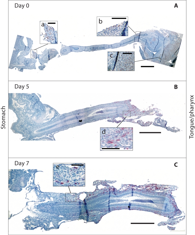
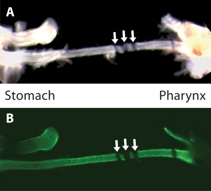
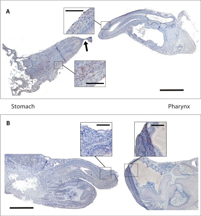
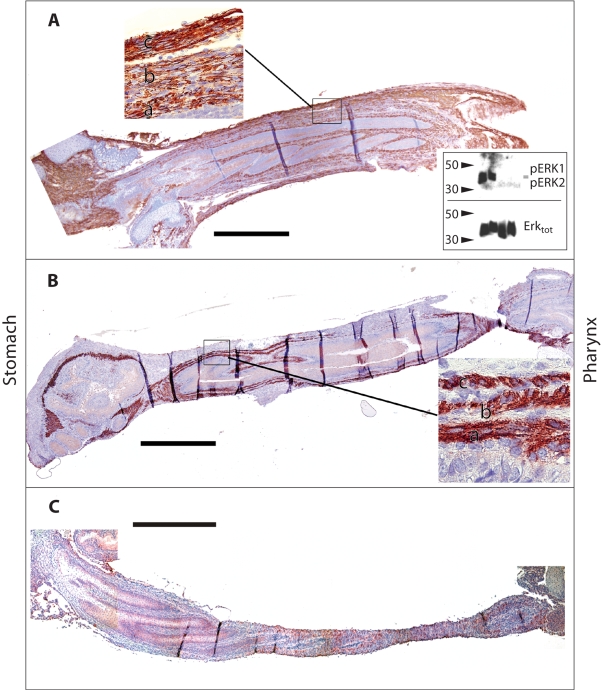
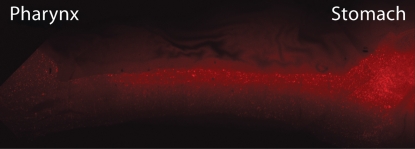
Esophageal striated myogenesis progresses differently from appendicular myogenesis, but the mechanism underlying this process is incompletely understood. Early theories of transdifferentiation of smooth muscle into striated muscle are not supported by transgenic fate-mapping experiments; however, the origin of esophageal striated muscle remains unknown. To better define the process of striated myogenesis, we examined myogenesis in murine fetal cultured esophageal whole-organ explants. Embryonic day 14.5 (E14.5) esophagi maintained a functional contractile phenotype for up to 7 days in culture. Striated myogenesis, as evidenced by myogenin expression, proceeded in a craniocaudal direction along the length of the esophagus. Esophageal length did not change during this process. Complete, but not partial, mechanical disruption of the rostral esophagus inhibited myogenesis distally. Addition of fibroblast growth factor-2 (FGF-2) to the culture media failed to inhibit striated myogenesis, but attenuated smooth muscle actin expression and reduced peristaltic activity. Inhibition of c-kit failed to inhibit peristalsis. These results suggest that striated myogenic precursors are resident along the entire length of the esophagus by day 14.5 and do not migrate along the esophagus after E14.5. Induction of myogenesis craniocaudally appears to require physical continuity of the esophagus and is not inhibited by FGF-2. Finally, peristalsis in E14.5 esophagi appears not to be regulated by interstitial cells of Cajal.
(c) 2008 S. Karger AG, Basel.
Figures
Similar articles
-
Smooth muscle persists in the muscularis externa of developing and adult mouse esophagus.J Muscle Res Cell Motil. 2007;28(2-3):153-65. doi: 10.1007/s10974-007-9112-y. Epub 2007 Jul 19. J Muscle Res Cell Motil. 2007. PMID: 17638088
-
Ultrastructural analysis of the smooth-to-striated transition zone in the developing mouse esophagus: emphasis on apoptosis of smooth and origin and differentiation of striated muscle cells.Dev Dyn. 2005 Jul;233(3):964-82. doi: 10.1002/dvdy.20436. Dev Dyn. 2005. PMID: 15918172
-
Effect of inter-swallow interval on striated esophagus peristalsis; a comparative study with smooth muscle esophagus.Neurogastroenterol Motil. 2023 Aug;35(8):e14608. doi: 10.1111/nmo.14608. Epub 2023 May 8. Neurogastroenterol Motil. 2023. PMID: 37154414 Free PMC article.
-
Embracing change: striated-for-smooth muscle replacement in esophagus development.Skelet Muscle. 2016 Aug 8;6:27. doi: 10.1186/s13395-016-0099-1. eCollection 2016. Skelet Muscle. 2016. PMID: 27504178 Free PMC article. Review.
-
Neuromuscular mechanisms of primary peristalsis.Am J Med. 1997 Nov 24;103(5A):40S-43S. doi: 10.1016/s0002-9343(97)00320-3. Am J Med. 1997. PMID: 9422621 Review.
Cited by
-
Molecular aspects of esophageal development.Ann N Y Acad Sci. 2011 Sep;1232:309-15. doi: 10.1111/j.1749-6632.2011.06071.x. Ann N Y Acad Sci. 2011. PMID: 21950820 Free PMC article.
-
Bi-layer silk fibroin grafts support functional tissue regeneration in a porcine model of onlay esophagoplasty.J Tissue Eng Regen Med. 2018 Feb;12(2):e894-e904. doi: 10.1002/term.2402. Epub 2017 Jun 20. J Tissue Eng Regen Med. 2018. PMID: 28084044 Free PMC article.
-
Oesophageal and sternohyal muscle fibres are novel Pax3-dependent migratory somite derivatives essential for ingestion.Development. 2013 Jul;140(14):2972-84. doi: 10.1242/dev.090050. Epub 2013 Jun 12. Development. 2013. PMID: 23760954 Free PMC article.
-
The expression of nestin delineates skeletal muscle differentiation in the developing rat esophagus.J Anat. 2011 Mar;218(3):311-23. doi: 10.1111/j.1469-7580.2010.01331.x. J Anat. 2011. PMID: 21323914 Free PMC article.
References
-
- Coffin J.D., Florkiewicz R.Z., Neumann J., Mort-Hopkins T., Dorn 2nd G.W., Lightfoot P., German R., Howles P.N., Kier A., O'Toole B.A. Abnormal bone growth and selective translational regulation in basic fibroblast growth factor (FGF-2) transgenic mice. Mol Biol Cell. 1995;6:1861–1873. - PMC - PubMed
-
- David S.G., Cebrian C., Vaughan E.D., Jr., Herzlinger D. c-kit and ureteral peristalsis. J Urol. 2005;173:292–295. - PubMed
-
- Jones N.C., Fedorov Y.V., Rosenthal R.S., Olwin B.B. ERK1/2 is required for myoblast proliferation but is dispensable for muscle gene expression and cell fusion. J Cell Physiol. 2001;186:104–115. - PubMed
-
- Kablar B., Tajbakhsh S., Rudnicki M.A. Transdifferentiation of esophageal smooth to skeletal muscle is myogenic bHLH factor-dependent. Development. 2000;127:1627–1639. - PubMed
-
- Kato S., Muraishi A., Miyamoto T., Fox J.C. Basic fibroblast growth factor regulates extracellular matrix and contractile protein expression independent of proliferation in vascular smooth muscle cells. In Vitro Cell Dev Biol Anim. 1998;34:341–346. - PubMed
Publication types
MeSH terms
Grants and funding
LinkOut - more resources
Full Text Sources
Other Literature Sources
