

OCT-guided hyaloid release for vitreomacular traction syndrome
- PMID: 18784444
- PMCID: PMC2629905
- DOI: 10.3341/kjo.2008.22.3.169
OCT-guided hyaloid release for vitreomacular traction syndrome
Abstract
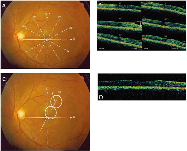
Purpose: To evaluate the usefulness of OCT retinal mapping in determining the configuration of a vitreomacular adhesion and selecting a meridian for entry into the subhyaloid space in patients with vitreomacular traction syndrome.
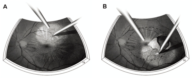
Methods: Six consecutive patients (6 eyes) with vitreomacular traction syndrome underwent vitrectomy with peeling of posterior hyaloid. Ocular coherence tomography (OCT) retinal mapping was performed preoperatively. Access to the subhyaloid space was made by creating an opening with a 25 gauge needle at a location where the detached posterior hyaloid membrane was farthest from the retinal surface. The location was selected based on six preoperative meridional OCT scans. The posterior hyaloid was then gently peeled off in a circular fashion around the fovea with a micropick. Visual acuity and foveal thicknesses were measured before the operation and 3 months afterwards.
Results: After the operation, visual acuity improved and central macular thicknesses were reduced significantly in all six patients. The best corrected visual acuity improved from 0.4 to 0.75 with a mean increase by 3.5 lines on a Snellen chart 3 months after the operation. The mean foveal thickness was reduced from 406 micrometer to 241 micrometer. The restoration of foveal pit was observed in five patients. Neither intraoperative nor postoperative complications were observed during the follow up period.
Conclusions: An OCT retinal mapping program is a valuable diagnostic tool in understanding the configuration of vitreomacular adhesion and planning the surgical approach for operating on vitreomacular traction syndrome.
Figures
References
-
- Reese AB, Jones IS, Cooper WC. Macular changes secondary to vitreous traction. Am J Ophthalmol. 1967;64:544–549. - PubMed
-
- Reese AB, Jones IS, Cooper WC. Vitreomacular traction syndrome confirmed histologically. Am J Ophthalmol. 1970;69:975–977. - PubMed
-
- Jaffe NS. Vitreous traction at the posterior pole of the fundus due to alteration in the vitreous posterior. Trans Am Acad Ophthalmol Otolaryngol. 1967;71:642–652. - PubMed
-
- Smiddy WE, Michels RG, Green WR. Morphology, pathology, and surgery of idiopathic vitreoretinal macular disorders. A review. Retina. 1990;10:288–296. - PubMed
-
- McDonald HR, Johnson RN, Schatz H. Surgical results in the vitreomacular traction syndrome. Ophthalmology. 1994;101:1397–1403. - PubMed
MeSH terms
LinkOut - more resources
Full Text Sources
Medical
