. 2009 Apr;34(5):1233-44.
doi: 10.1038/npp.2008.143. Epub 2008 Sep 10.

Ethanol enhances glutamate transmission by retrograde dopamine signaling in a postsynaptic neuron/synaptic bouton preparation from the ventral tegmental area

Affiliations

Ethanol enhances glutamate transmission by retrograde dopamine signaling in a postsynaptic neuron/synaptic bouton preparation from the ventral tegmental area

Chunyu Deng et al. Neuropsychopharmacology. 2009 Apr.

Abstract

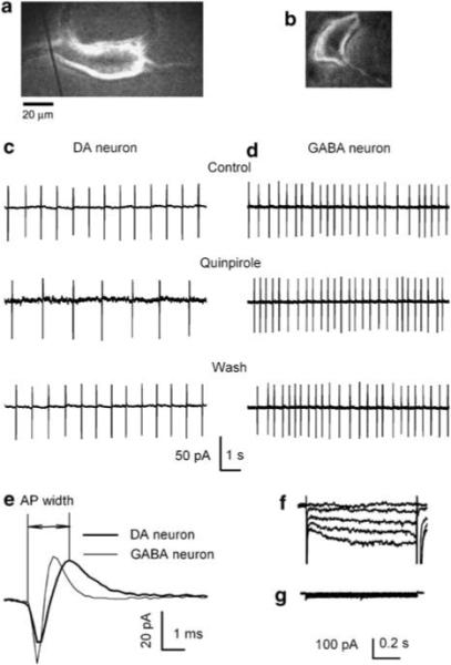
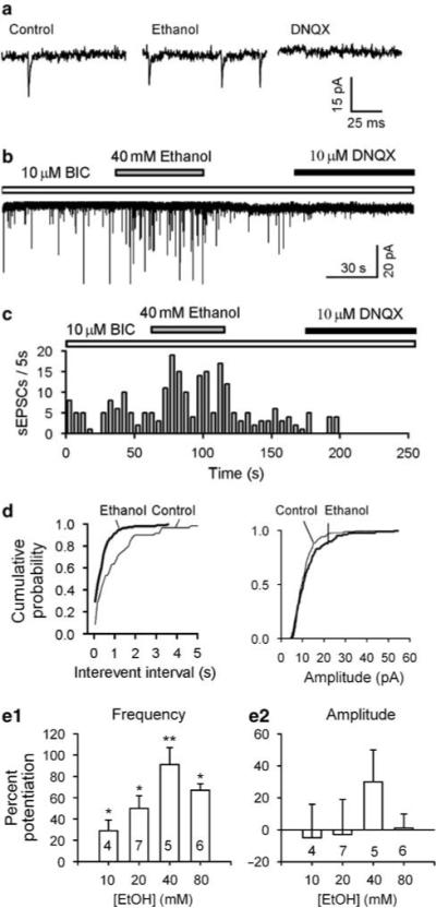
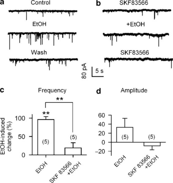
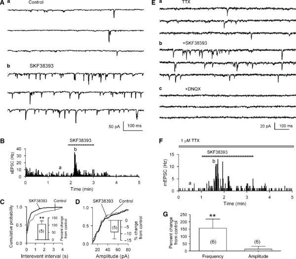
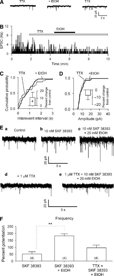
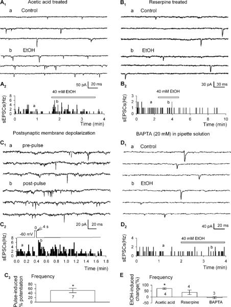
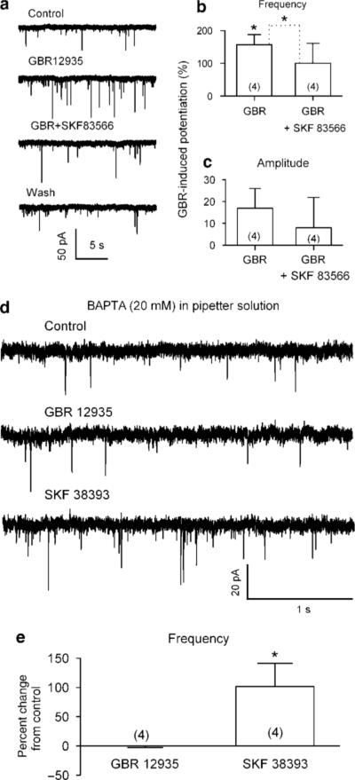
It is well documented that somatodendritically released dopamine is important in the excitability and synaptic transmission of midbrain dopaminergic neurons. Recently we showed that in midbrain slices, acute ethanol exposure facilitates glutamatergic transmission onto dopaminergic neurons in the ventral tegmental area (VTA). The VTA is a brain region critical to the rewarding effects of abused drugs, including ethanol. We hypothesized that ethanol facilitation might result from an increase in somatodendritically released dopamine, which acts retrogradely on dopamine D(1) receptors on glutamate-releasing axons and consequently leads to an increase in glutamate release onto dopaminergic neurons. To further test this hypothesis and to examine whether ethanol facilitation can occur at the single-cell level, VTA neurons were freshly isolated from rat brains using an enzyme-free procedure. These isolated neurons retain functional synaptic terminals, including those that release glutamate. Spontaneous excitatory postsynaptic currents (sEPSCs) mediated by glutamate alpha-amino-3-hydroxy-5-methylisoxazole-4-propionic acid receptors were recorded from these freshly isolated putative dopaminergic neurons. We found that acute application of clinically relevant concentrations of ethanol (10-80 mM) significantly facilitated the frequency of sEPSCs but not their mean amplitude. Ethanol facilitation was mimicked by the D(1) agonist SKF 38393 and by the dopamine uptake blocker GBR 12935 but was blocked by the D(1) antagonist SKF 83566, and by depleting dopamine stores with reserpine, as well as by chelating postsynaptic calcium with BAPTA. Furthermore, the sodium channel blocker tetrodotoxin eliminated the facilitation of sEPSCs induced by ethanol but not by SKF 38393. These results constitute the first evidence from single isolated cells of ethanol facilitation of glutamate transmission to dopaminergic neurons in the VTA. In addition, we show that ethanol facilitation has a postsynaptic origin and a presynaptic locus. Furthermore, ethanol stimulation of a single dopaminergic neuron is capable of eliciting the release of somatodendritic dopamine, which is sufficient to influence glutamatergic transmission at individual synapses.


Figures

Figure 1
Figure 1
Identification of DAergic neurons. Photomicrograph of a mechanically dissociated putative DAergic neuron (a) and a putative GABAergic neuron (b). The much reduced dendritic arbors of such cells facilitate space clamp. Spontaneous firings recorded with loose-patch cell-attached mode showing the inhibitory response of the DAergic neuron (c) and the no response of the GABAergic neuron (d) to the D2/3 receptor antagonist, quinpirole. (e) Comparison of single spontaneous action potentials recorded from a putative DAergic neuron (thicker line, with a half-width of 1.7 ms) and a putative GABAegic neuron (thinner line, with a half-width of 1.1 ms). Under voltage clamp, the currents induced in such a DAergic cell by a series of hyperpolarizing voltage pulses (the 10 mV steps to −110 mV) show the prominent Ih of the DAergic (f) but not in the GABAergic (g) neuron.
Figure 2
Figure 2
Ethanol enhances frequency of spontaneous excitatory postsynaptic currents (sEPSCs) recorded from mechanically dissociated DAergic neurons. sEPSCs were recorded in the presence of bicuculline (10 μM) at a VH of −65 mV. The frequency of sEPSCs was increased by 40 mM ethanol and the sEPSCs were eliminated by 10 −M DNQX (a–c). (c) Time course of ethanol-induced enhancement of sEPSC frequency in one experiment. (d) Cumulative probability of sEPSC interevent intervals (left panel) and current amplitudes (right panel) of the same data. (e) Dose-dependent potentiation of sEPSC frequency (e1), but unaltered amplitude (e2; mean ± SEM, number of cells in each group is indicated). *p<0.05, **p<0.01, paired t-test for ethanol application vs pre-ethanol control.
Figure 3
Figure 3
Effect of the D1R antagonist on ethanol facilitation of sEPSCs. (a) Current traces show that ethanol enhancement of sEPSCs was eliminated by 10 μM SKF 83566 (D1R antagonist; b). Summary of ethanol-induced changes (%) in sEPSC frequency (c) and amplitude (d) in the absence (EtOH) and the presence of SKF 83566 (SKF 83566 + EtOH, data from six cells). **p<0.01, paired t-test for ethanol application vs preethanol control.
Figure 4
Figure 4
Effect of the D1R agonist on sEPSCs and mEPSCs in isolated neurons. (A) The D1R agonist SKF 38393 (10 μM) increased sEPSC frequency in a putative DAergic neuron in the VTA. (B) Time course of changes induced by SKF 38393. The traces in (A) were taken at times indicated by (a) and (b). The change in frequency is also demonstrated by the much increased incidence of shorter intervals between sEPSCs. A representative cumulative probability plot of interevent interval (C), but there was no associated change in sEPSC amplitudes (D). The inset shows mean ± SEM of % changes in sEPSC frequency (C) and amplitude (D) induced by SKF 38393 (10 μM; n = 5). **p<0.01, paired t-test for SKF 38393 application vs pre-SKF 38393. (E) SKF 38393 (10 μM) increased mEPSC frequency in the presence of TTX (1 μM) in a putative DAergic neuron in the VTA. (F) Time course of changes induced by SKF 38393. The traces in (E) were taken at times indicated by (a) and (b). (G) Summary of results (mean ± SEM of % changes) from six cells showing that SKF 38393 (10 μM) significantly increased mEPSC frequency (left column) but not the amplitude (right column). **p<0.01, paired t-test for SKF 38393 application vs pre-SKF 38393.
Figure 5
Figure 5
Ethanol has no effect on mEPSCs. The mEPSCs were recorded in VTA DAergic neurons at a VH of −65 mV, in the presence of bicuculline (10 μM) and tetrodotoxin (TTX, 1 μM). (A) Application of 40 mM ethanol did not change the incidence of mEPSCs. (B) Time course of ethanol-induced changes of sEPSC frequency in one experiment. (C, D) Representative cumulative probability plots of intervals between mEPSCs (C) and their amplitudes (D) remained unchanged. Inset: mean data (± SEM, from four neurons) confirm absence of significant effect of ethanol on frequency and amplitude of mEPSCs. (E) Ethanol (20 mM) increased the enhancement of sEPSC frequency induced by 20 nM SKF 38393 (c) but not in the presence (e) of TTX (1 μM). (F) Mean data (± SEM, from four neurons) confirm ethanol (20 mM) increased 20 nM SKF 38393-induced enhancement of sEPSC frequency in the absence but not in the presence of TTX (1 μM). **p<0.01, t-test for SKF 38393 application vs SKF 38393 + ethanol.
Figure 6
Figure 6
The release of dopamine is necessary for ethanol's facilitation of sEPSCs. (A1, B1) Current traces show that ethanol enhancement of sEPSCs was observed in neurons from slices pretreated with acetic acid (A1), but not from reserpine (B1). (A2, B2) Time course of ethanol-induced change of sEPSC frequency in one experiment. The traces in A1 and B1 were taken at times indicated by (a) and (b). (C1) Current traces show that a 4 s depolarization pulse (from −60 to 0 mV) increased sEPSC frequency. (C2) Time course of depolarization-induced changes of sEPSC frequency in one experiment. (C3) Mean data (± SEM, from seven neurons) show depolarization induced potentiation of sEPSC frequency. *p<0.05, paired t-test for postpulse vs prepulse control. (D1) Current traces show that 40 mM ethanol did not change the sEPSCs when the pipette solution contained 20 mM BAPTA. (D2) Time course of 40 mM ethanol-induced changes of sEPSC frequency in one experiment in which BAPTA was in the pipette solution. (E) Mean data (± SEM) show changes in sEPSC frequency induced by 40 mM ethanol under different experimental conditions. *p<0.05, paired t-test for ethanol application vs pre-ethanol control, or as indicated, nonpaired t-test for reserpine group vs reserpine + ethanol group.
Figure 7
Figure 7
Ethanol-induced increase in sEPSC frequency is mimicked by blocking dopamine transport. (a) Traces illustrating increase in sEPSC frequency produced by GBR 12935 (1 μM, a dopamine transport blocker), which is blocked by the D1R antagonist SKF 83566 (10 μM, n = 4). (b) Summary of GBR 12935-induced changes in sEPSC frequency, which was blocked by SKF 83566 (data from four cells). *p<0.05, paired t-test for GBR 12935 application vs pre-GBR 12935, or as indicated for GBR 12935 group vs GBR 12935 + SKF 83566. (c) No change in amplitude was seen in GBR 12935 either in the absence or the presence of 10 μM SKF 83566. (d) Traces illustrating increase in sEPSC frequency produced by 10 μM SKF 38393 but not by 1 μM GBR 12935 when 20 mM BAPTA was in the pipette solution. (e) Mean data (± SEM, from four neurons) show that SKF 38393, but not GBR 12935, increased sEPSC frequency when pipette solution contained 20 mM BAPTA. *p<0.05, paired t-test for SKF 38393 application vs pre-SKF 38393.

References

    1. Abarca J, Gysling K, Roth RH, Bustos G. Changes in extracellular levels of glutamate and aspartate in rat substantia nigra induced by dopamine receptor ligands: in vivo micro-dialysis studies. Neurochem Res. 1995;20:159–169. - PubMed
    1. Adell A, Artigas F. The somatodendritic release of dopamine in the ventral tegmental area and its regulation by afferent transmitter systems. Neurosci Biobehav Rev. 2004;28:415–431. - PubMed
    1. Aghajanian GK, Bunney BS. Pharmacological characterization of dopamine ‘autoreceptors’ by microiontophoretic single-cell recording studies. Adv Biochem Psychopharmacol. 1977;16:433–438. - PubMed
    1. Akaike N, Moorhouse AJ. Techniques: applications of the nerve-bouton preparation in neuropharmacology. Trends Pharmacol Sci. 2003;24:44–47. - PubMed
    1. Alberto CO, Trask RB, Quinlan ME, Hirasawa M. Bidirectional dopaminergic modulation of excitatory synaptic transmission in orexin neurons. J Neurosci. 2006;26:10043–10050. - PMC - PubMed

Publication types

MeSH terms