. 2008 Oct 8;27(19):2471-83.
doi: 10.1038/emboj.2008.182. Epub 2008 Sep 11.

LIS1 and NDEL1 coordinate the plus-end-directed transport of cytoplasmic dynein

Affiliations

LIS1 and NDEL1 coordinate the plus-end-directed transport of cytoplasmic dynein

Masami Yamada et al. EMBO J. .

Abstract

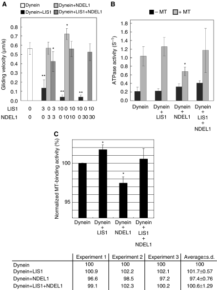
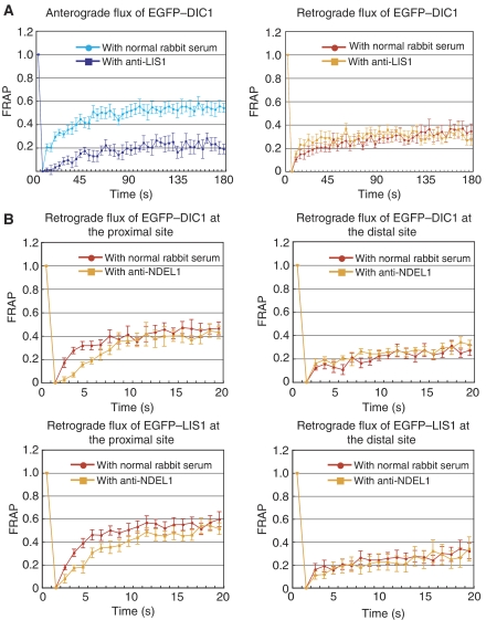
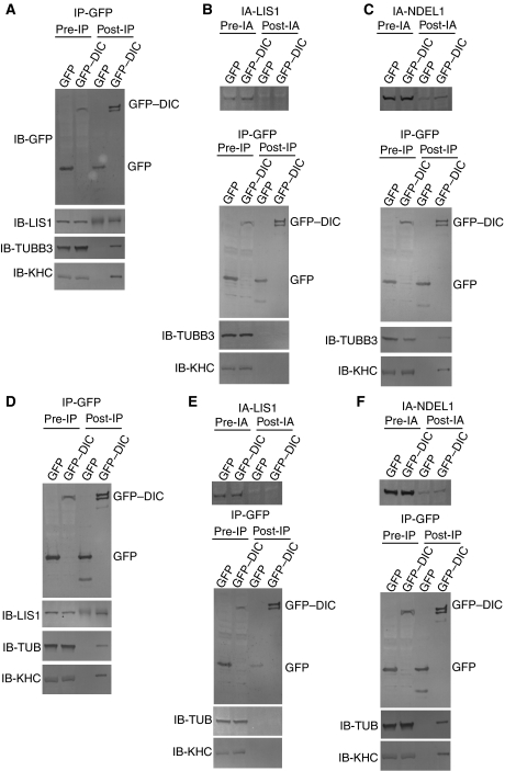
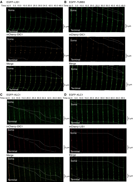
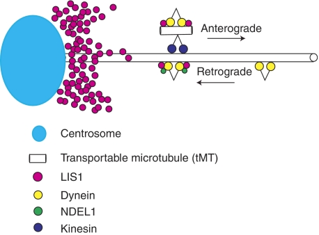
LIS1 was first identified as a gene mutated in human classical lissencephaly sequence. LIS1 is required for dynein activity, but the underlying mechanism is poorly understood. Here, we demonstrate that LIS1 suppresses the motility of cytoplasmic dynein on microtubules (MTs), whereas NDEL1 releases the blocking effect of LIS1 on cytoplasmic dynein. We demonstrate that LIS1, cytoplasmic dynein and MT fragments co-migrate anterogradely. When LIS1 function was suppressed by a blocking antibody, anterograde movement of cytoplasmic dynein was severely impaired. Immunoprecipitation assay indicated that cytoplasmic dynein forms a complex with LIS1, tubulins and kinesin-1. In contrast, immunoabsorption of LIS1 resulted in disappearance of co-precipitated tubulins and kinesin. Thus, we propose a novel model of the regulation of cytoplasmic dynein by LIS1, in which LIS1 mediates anterograde transport of cytoplasmic dynein to the plus end of cytoskeletal MTs as a dynein-LIS1 complex on transportable MTs, which is a possibility supported by our data.


Figures

Figure 1
Figure 1
Effects of LIS1/NDEL1 on the in vitro motor properties of cytoplasmic dynein. (A) Dependence of gliding velocity of microtubules (MTs) on the concentration of LIS1 and NDEL1. Molecular ratio is indicated at the bottom. Note: LIS1 displayed dose-dependent inhibition of dynein motility, whereas NDEL1 facilitated dissociation of dynein with MTs. The presence of LIS1 and NDEL1 restored dynein binding with MTs. No translocation was counted due to complete dissociation of dynein at the highest concentration of NDEL1. The P-value was calculated using a Student's t-test (*P<0.05, **P<0.01). (B) MgATPase activities of cytoplasmic dynein. Each protein was added at a 10-fold stoichiometric amount to the cytoplasmic dynein heavy chain. Left bars: activity without MTs. Right bars: calculated kcat for each protein combination. The P-value was calculated using a Student's t-test (*P<0.05). (C) MT-binding assay. The amount of cytoplasmic dynein bound to MTs was indicated in percentage (and s.d.) of total cytoplasmic dynein. LIS1 slightly increased cytoplasmic dynein binding to MTs, whereas NDEL1 reduced the cytoplasmic dynein binding. When both LIS1 and NDEL1 were present, the reduced binding is restored to the level of MTs with LIS1 alone. Three independent experiments were performed, and we found significant difference. Intensity of the band of SDS–PAGE was measured and was normalized as shown at the bottom. The P-value was calculated using a Student's t-test (*P<0.05).
Figure 2
Figure 2
FRAP analysis of dorsal root ganglia (DRG) neuron. FRAP analysis of DRG neuron expressing EGFP-tagged proteins. The graph shows the relative fluorescent recovery directly after bleaching. The prebleach level is normalized to 1.0. (A) Dynein recovery in the presence of an anti-LIS1 blocking antibody. (B) Retrograde dynein and LIS1 recovery in the presence of AMPPNP and an anti-NDEL1 blocking antibody.
Figure 3
Figure 3
Immunoprecipitation and immunoabsorption assay. (A) Total cell extracts from DRG neurons transiently transfected with either a pGFP vector encoding DIC1 or an empty pGFP vector were immunoprecipitated with an anti-GFP antibody. The input (5%) (pre-IP) and the immunoprecipitates (post-IP) were analysed by SDS–PAGE and western blot with antibodies against the proteins is indicated on the left. Note: LIS1, TUBB3 and KHC1 are precipitated with GFP–DIC1, whereas the GFP control did not result in any precipitation of these proteins. IB with an anti-LIS1 antibody for the GFP control displayed some background due to cross reactivity of the same rabbit serum used rather than any specific signal. (B) Immunoabsorption assay to remove endogenous LIS1 by an anti-LIS1 antibody. Upper panel: total cell extracts from DRG neurons were immunoabsorbed with an anti-LIS1 antibody. The input (5%) (pre-IA) and the post-immunoabsorption (post-IA) were analysed by SDS–PAGE and western blot. Note: immunoabsorption efficiently removed endogenous LIS1 from the total extracts. Immunoprecipitation assay by an anti-GFP antibody. Lower panels: LIS1-immunoabsorbed cell extracts from DRG neurons were immunoprecipitated by anti-GFP antibodies. The input (5%) (pre-IP) and the immunoprecipitates (post-IP) were analysed by SDS–PAGE and western blot with antibodies against the proteins indicated on the left. Note: immunoabsorption of LIS1 resulted in the absence of TUBB3 and KHC co-precipitation with GFP–DIC1. (C) Immunoabsorption assay to remove endogenous NDEL1 by an anti-NDEL1 antibody. Upper panel: total cell extracts from DRG neurons were immunoabsorbed with an anti-NDEL1 antibody. The input (5%) (pre-IA) and the immunoabsorption (post-IA) were analysed by SDS–PAGE and western blot. Note: immunoabsorption efficiently removed endogenous NDEL1 from the total extracts. Immunoprecipitation assay by an anti-GFP antibody. Lower panels: NDEL1-immunoabsorbed cell extracts from DRG neurons were immunoprecipitated by anti-GFP antibodies. The input (5%) (pre-IP) and the immunoprecipitates (post-IP) were analysed by SDS–PAGE and western blot with antibodies against the proteins indicated on the left. Note: TUBB3 and KHC co-precipitates with GFP–DIC1 in the absence of NDEL1. (DF) MEF cells were transfected with GFP–DIC1 or GFP control plasmid, followed by immunoprecipitation using an anti-GFP antibody. The same immunoprecipitation and immunoabsorption/immunoprecipitation assays were applied to MEF cells, and reproducible results were obtained. Note: in these series of experiments, we used GFP–TUBB5 and an anti-tubulin β antibody instead of GFP–TUBB3 and an anti-tubulin β3 antibody.
Figure 4
Figure 4
Imaging of movements of LIS1, TUBB3, DIC1 and KLC1 in DRG. Direct visualization of the anterograde movement of fluorescence-tagged proteins in DRG neurons using confocal microscopy. (A) EGFP–LIS1 and mCherry–DIC1. (B) EGFP–TUBB3 and mCherry–DIC1. (C) EGFP–KLC1 and mCherry–DIC. (D) EGFP–KLC1 and mCherry–LIS1. Dotted lines indicate dynamic colocalization. Yellow signal indicates colocalized transport. Calculated speed of particles was distributed in 0.5–1.6 μm/s, consistent with the speed of kinesin.
Figure 5
Figure 5
Model of dynein regulation by LIS1 and NDEL1. Schematic presentation of the dynein regulation by LIS1 and NDEL1. LIS1 fixes dynein on tMT (freighter tubulins). This complex is transported to the plus end of cytoskeletal MTs in a kinesin-dependent manner. In contrast, NDEL1 activates the dynein–LIS1 complex, and enables LIS1 protein to accumulate around the centrosome by dynein-dependent transport. Presumably, the generation of the dynein–LIS1–tMT freighter complex requires high concentrations of LIS1 protein around the centrosome.

Similar articles

Cited by

References

    1. Bingham JB, King SJ, Schroer TA (1998) Purification of dynactin and dynein from brain tissue. Methods Enzymol 298: 171–184 - PubMed
    1. Bouchard P, Penningroth SM, Cheung A, Gagnon C, Bardin CW (1981) erythro-9-[3-(2-Hydroxynonyl)]adenine is an inhibitor of sperm motility that blocks dynein ATPase and protein carboxylmethylase activities. Proc Natl Acad Sci USA 78: 1033–1036 - PMC - PubMed
    1. Dobyns WB, Curry CJ, Hoyme HE, Turlington L, Ledbetter DH (1991) Clinical and molecular diagnosis of Miller–Dieker syndrome. Am J Hum Genet 48: 584–594 - PMC - PubMed
    1. Dobyns WB, Reiner O, Carrozzo R, Ledbetter DH (1993) Lissencephaly. A human brain malformation associated with deletion of the LIS1 gene located at chromosome 17p13. JAMA 270: 2838–2842 - PubMed
    1. Dujardin DL, Barnhart LE, Stehman SA, Gomes ER, Gundersen GG, Vallee RB (2003) A role for cytoplasmic dynein and LIS1 in directed cell movement. J Cell Biol 163: 1205–1211 - PMC - PubMed

Publication types

MeSH terms