Review
. 2008 Oct 8;27(19):2457-70.
doi: 10.1038/emboj.2008.169. Epub 2008 Sep 11.

Function and dysfunction of the PI system in membrane trafficking

Affiliations
Review

Function and dysfunction of the PI system in membrane trafficking

Mariella Vicinanza et al. EMBO J. .

Abstract

The phosphoinositides (PIs) function as efficient and finely tuned switches that control the assembly-disassembly cycles of complex molecular machineries with key roles in membrane trafficking. This important role of the PIs is mainly due to their versatile nature, which is in turn determined by their fast metabolic interconversions. PIs can be tightly regulated both spatially and temporally through the many PI kinases (PIKs) and phosphatases that are distributed throughout the different intracellular compartments. In spite of the enormous progress made in the past 20 years towards the definition of the molecular details of PI-protein interactions and of the regulatory mechanisms of the individual PIKs and phosphatases, important issues concerning the general principles of the organisation of the PI system and the coordination of the different PI-metabolising enzymes remain to be addressed. The answers should come from applying a systems biology approach to the study of the PI system, through the integration of analyses of the protein interaction data of the PI enzymes and the PI targets with those of the 'phenomes' of the genetic diseases that involve these PI-metabolising enzymes.


Figures
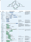
Figure 1
Figure 1
The PI kinases and phosphatases and their genetic defects. (A) Schematic representation of the PI metabolic cycle with the PIKs indicated in blue, and the PI phosphatases in green. (B) Listing of the different isoforms of the PIKs and PI phosphatases, together with their domain organisation and their corresponding genetic disease and knockout or knockdown phenotypes in mice. RSBD, regulatory subunit-binding domain; C2, conserved region 2; HD, helical domain; PRD, proline-rich domain, NLS, nuclear localisation signal; LKU, lipid kinase unique domain; PH, pleckstrin-homology domain; SRD, serine-rich domain; CRD, cysteine-rich domain; FYVE, Fab1 YOTB Vac1 EEA1; DEP, domain present in dishevelled, EGL-10 and pleckstrin; TCP1, tailless complex polypeptide-1; SPEC, spectrin repeat; PH-GRAM, pleckstrin homology glucosyltransferases, Rab-like GTPase activators and myotubularins; DENN, differentially expressed in normal versus neoplastic; LZ, leucine zipper; TM, transmembrane domain; ASH, abnormal spindle-like microcephaly-associated protein (ASPM), C. elegans centrosomal protein (SPD-2), hydrocephalus-associated protein (Hydin); RhoGAP, Rho-GTPase-activating protein; SAC, yeast suppressor of actin 1; Skitch, SKIP carboxyl homology domain; SH2, phosphotyrosine-binding module 2; SAM, sterile alpha-motif domain.
Figure 2
Figure 2
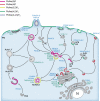
Subcellular distribution of the PIs and PI-metabolising enzymes. The localisation of the different PIKs (blue) and PI phosphatases (green), as well as the predominant PI species (as visualised by PI-binding protein domains) in the different cell compartments. It is noted that many of the PI-metabolising enzymes are present in more than one cellular compartment, and their overall distributions do not completely fit with the PI map, as indicated using the PI-binding protein probes (see text for details). The PIKs and PI phosphatases are indicated according to the nomenclature given in Figure 1. PM, plasma membrane; EE, early endosome; SE: sorting endosomes; RE, recycling endosome; LY, lysosome; MVB/LE, multivesicular body/late endosome; PAS, pre-autophagosomal structure; PH, phagosome; TGN, trans-Golgi network; GC, Golgi complex; ER: endoplasmic reticulum; N, nucleus.
Figure 3
Figure 3
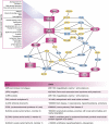
Co-expression network of the PI-metabolising enzymes. Co-expression network of the PI-metabolising enzymes (PIKs, blue ellipses; PI phosphatases, green ellipses; as listed in Figure 1), focused on the 3- and 5-phosphatases. The analysis was performed using the COXPRESdb database of gene expression profiles from a variety of normal and pathological conditions (from a total of 123 experiments; Obayashi et al, 2008 no. 22). Here, using the 3- and 5-phosphatase genes as individual queries and analysing the most significantly co-expressed genes (according to the weighted Pearson's correlation coefficient between gene probes, and choosing 0.4 as a threshold; Obayashi et al 2008), we selected specifically for the genes for PI-metabolising enzymes. Groups of genes extensively connected in the network represent co-expressed gene clusters that are likely to be involved in common cell functions. The 3- and 5-phosphatase genes responsible for genetic diseases (see Figure 1) are highlighted in bold. Four interconnected enzyme clusters emerge from the analysis, each containing a disease gene (see Figure 1). In particular, OCRL gene is part of a cluster that also comprises PIP5K1A, INPP5A and MTMR1.
Figure 4
Figure 4
Interaction map of the products of the genes responsible or candidate for disorders of kidney proximal tubule cells (PTCs). The genes responsible or strong candidate for diseases due to dysfunction of kidney PTCs are shown (large red ellipses), together with selected interactors, including genes for which the knock out causes proteinuria and tubular acidosis in mice (rectangles). The interaction map was generated using Osprey (powered by Human GRID, General Repository of Interaction Datasets). The components of the network that are active on or sensitive to PI45P2 (the substrate of the 5-phosphatase OCRL) are marked in yellow. LRP2 encodes for megalin, a multiligand receptor that, together with cubilin (CBN), is responsible for the resorption of the indicated LMW proteins by PTCs (Igarashi et al, 2002; Christensen and Gburek, 2004). ARH, autosomal recessive hypercholesterolaemia; DAB2, disabled homologue 2; GIPC, GAIP C terminus interacting protein; PIP5K1C, phosphatidylinositol-4-phosphate 5-kinase type I gamma; SYNJ2BP, synaptojanin 2-binding protein; CLTC, clathrin heavy chain 1; AP2M1, clathrin coat assembly protein AP50, Myo VI, myosin 6, APPL1, adapter protein containing PH domain, PTB domain and leucine zipper motif 1; Arf6, ADP ribosylation factor 6; PIP5KL1, phosphatidylinositol-4-phosphate 5-kinase-like 1; SYNJ2, synaptojanin-2; RAC1, Ras-related C3 botulinum toxin substrate 1; RhoA, transforming protein RhoA; IP3R, inositol 1,4,5-trisphosphate receptor type 1; AHCYL1, putative adenosylhomocysteinase 2; SLC9A1, Na/H exchanger 1 (NHE-1) (solute carrier family 9 member 1); ARHGDIA, Rho GDP dissociation inhibitor 1 (Rho GDI 1); ARHGDIB, Rho GDP dissociation inhibitor 2 (Rho GDI 2); PIK3R1, phosphatidylinositol 3-kinase regulatory alpha subunit; EZR, Ezrin; SLC4A8, solute carrier family 4, sodium bicarbonate cotransporter, member 8; SLC9A3R1, Na(+)/H(+) exchange regulatory cofactor NHE-RF) (NHERF-1); SLC9A3R2, Na(+)/H(+) exchange regulatory cofactor NHE-RF2 (NHERF-2); PLCB1, phospholipase C-beta-1; ACTA1, alpha actin 1; ACTC, alpha cardiac actin.

Similar articles

Cited by

References

    1. Aoyagi K, Sugaya T, Umeda M, Yamamoto S, Terakawa S, Takahashi M (2005) The activation of exocytotic sites by the formation of phosphatidylinositol 4,5-bisphosphate microdomains at syntaxin clusters. J Biol Chem 280: 17346–17352 - PubMed
    1. Attree O, Olivos IM, Okabe I, Bailey LC, Nelson DL, Lewis RA, McInnes RR, Nussbaum RL (1992) The Lowe's oculocerebrorenal syndrome gene encodes a protein highly homologous to inositol polyphosphate-5-phosphatase. Nature 358: 239–242 - PubMed
    1. Bai J, Chapman ER (2003) Application of fluorescent probes to study mechanics and dynamics of Ca2+-triggered synaptotagmin C2 domain–membrane interactions. Methods Enzymol 360: 238–258 - PubMed
    1. Balla A, Kim YJ, Varnai P, Szentpetery Z, Knight Z, Shokat KM, Balla T (2008) Maintenance of hormone-sensitive phosphoinositide pools in the plasma membrane requires phosphatidylinositol 4-kinase III{alpha}. Mol Biol Cell 19: 711–721 - PMC - PubMed
    1. Behnia R, Munro S (2005) Organelle identity and the signposts for membrane traffic. Nature 438: 597–604 - PubMed

Publication types

MeSH terms

Substances