

Fulminant EBV-driven CD8 T-cell lymphoproliferative disorder following primary acute EBV infection: a unique spectrum of T-cell malignancy
- PMID: 18784807
- PMCID: PMC2480557
Fulminant EBV-driven CD8 T-cell lymphoproliferative disorder following primary acute EBV infection: a unique spectrum of T-cell malignancy
Abstract
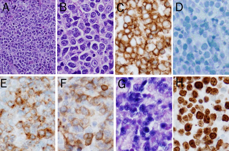
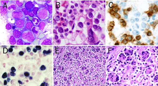
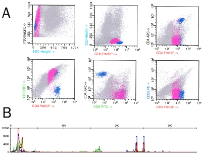
Fulminant Epstein-Barr virus (EBV)-driven clonal T-cell lymphoproliferative disorder (T-LPD) is rare and most patients are of Asian origin. The disease usually develops shortly after primary acute EBV infection and the mechanism remains poorly understood. Here we report such a rare case in a 28-year-old Caucasian female with systemic lupus erythematosus (SLE). Immunophenotypic and molecular studies revealed that the proliferating lymphoid cells displayed a CD8(+) T-cell phenotype with clonal rearrangement of the T-cell receptor gamma gene. Epstein-Barr virus-encoded RNA was also observed in the clonal lymphoid cells by in situ hybridization. The patient subsequently developed fatal virus-associated hemophagocytic syndrome one month after the primary acute EBV infection. The case represents the first report of fulminant EBV-driven CD8(+) T-LPD occurring in an immunocompromised Caucasian SLE patient. This study, along with studies of similar Asian cases reported in the literature, suggests that dysregulated immunity due to either acquired or genetically determined susceptibility may result in an abnormal response to primary EBV infection and contribute to the pathogenesis of EBV-mediated fatal T-LPD.
Keywords: Epstein-Barr virus; Fatal infectious mononuleosis; T-cell lymphoproliferative disorder; hemophagocytosis; systemic lupus erythematosus; virus-associated hemophagocytic syndrome.
Figures
Similar articles
-
Fulminant EBV(+) T-cell lymphoproliferative disorder following acute/chronic EBV infection: a distinct clinicopathologic syndrome.Blood. 2000 Jul 15;96(2):443-51. Blood. 2000. PMID: 10887104
-
Systemic Epstein-Barr Virus-Positive T/NK Lymphoproliferative Diseases With SH2D1A/XIAP Hypomorphic Gene Variants.Front Pediatr. 2019 May 21;7:183. doi: 10.3389/fped.2019.00183. eCollection 2019. Front Pediatr. 2019. PMID: 31231620 Free PMC article.
-
The ambiguous boundary between EBV-related hemophagocytic lymphohistiocytosis and systemic EBV-driven T cell lymphoproliferative disorder.Int J Clin Exp Pathol. 2014 Aug 15;7(9):5738-49. eCollection 2014. Int J Clin Exp Pathol. 2014. PMID: 25337215 Free PMC article. Review.
-
Systemic Epstein-Barr virus-positive T-cell lymphoproliferative disorders of childhood with fulminant leukocytosis and tumor lysis: a case report with autopsy findings.Int J Hematol. 2018 Jul;108(1):112-117. doi: 10.1007/s12185-017-2380-4. Epub 2017 Dec 7. Int J Hematol. 2018. PMID: 29218495
-
Epstein-Barr virus-associated T/natural killer-cell lymphoproliferative disorders.J Dermatol. 2014 Jan;41(1):29-39. doi: 10.1111/1346-8138.12322. J Dermatol. 2014. PMID: 24438142 Review.
Cited by
-
Successful treatment of immediate allogeneic myeloablative hematopoietic stem cell transplantation from a HLA-mismatched sibling donor for active systemic epstein-barr virus-positive T-cell lymphoproliferative disease of childhood following primary acute epstein-barr virus infection.Clin Case Rep. 2015 Apr;3(4):231-6. doi: 10.1002/ccr3.204. Epub 2015 Feb 2. Clin Case Rep. 2015. PMID: 25914814 Free PMC article.
-
Epstein-Barr virus: general factors, virus-related diseases and measurement of viral load after transplant.Rev Bras Hematol Hemoter. 2011;33(5):383-8. doi: 10.5581/1516-8484.20110103. Rev Bras Hematol Hemoter. 2011. PMID: 23049344 Free PMC article.
-
Sequential monitoring of serum IL-6, TNF-α, and IFN-γ levels in a CAEBV patient treated by plasma exchange and immunochemotherapy.Int J Hematol. 2012 Nov;96(5):669-73. doi: 10.1007/s12185-012-1170-2. Epub 2012 Sep 16. Int J Hematol. 2012. PMID: 22983646
-
Infectious mononucleosis complicated by transitory Epstein-Barr virus infection of T and natural killer cells.J Hematop. 2024 Sep;17(3):129-137. doi: 10.1007/s12308-024-00595-6. Epub 2024 Jul 5. J Hematop. 2024. PMID: 38967898 Free PMC article.
-
Epstein-Barr Virus-Associated T and NK-Cell Lymphoproliferative Diseases.Front Pediatr. 2019 Mar 15;7:71. doi: 10.3389/fped.2019.00071. eCollection 2019. Front Pediatr. 2019. PMID: 30931288 Free PMC article. Review.
References
-
- Straus SE, Cohen JI, Tosato G, Meier J. Epstein-Barr virus infections: biology, pathogenesis and management. Ann Int Med. 1993;118:45–58. - PubMed
-
- Auwaerter PG. Recent advances in the understanding of infectious mononucleosis: are prospects improved for treatment or control? Expert Rev Anti Infect Ther. 2006;4:1039–1049. - PubMed
-
- Weisenburger DD, Purtilo DT. Failure in immunological control of the virus infection: fatal infectious mononucleosis. In: Epstein MA, Achong BG, editors. The Epstein-Barr Virus: Recent Advances. London, England: Heinmann Medical Books; 1986. pp. 129–161.
-
- Coffey AJ, Brooksbank RA, Brandau O, Oohashi T, Howell GR, Bye JM, Cahn AP, Durham J, Heath P, Wray P, Pavitt R, Wilkinson J, Leversha M, Huckle E, Shaw-Smith CJ, Dunham A, Rhodes S, Schuster V, Porta G, Yin L, Serafini P, Sylla B, Zollo M, Franco B, Bolino A, Seri M, Lanyi A, Davis JR, Webster D, Harris A, Lenoir G, De St Basile G, Jones A, Behloradsky BH, Achatz H, Murken J, Fassler R, Sumegi J, Romeo G, Vaudin M, Ross MT, Meindl A, Bentley DR. Host response to EBV infection in X-linked lymphoproliferative disease results from mutations in an SH2-domain encoding gene. Nat Genet. 1998;20:129–135. - PubMed
-
- Mroczek EC, Weisenburger DD, Grierson H, Markin R, Purtilo DT. Fatal infectious mononucleosis and virus-associated hemophagocytic syndrome. Arch Pathol Lab Med. 1987;111:530–535. - PubMed
LinkOut - more resources
Full Text Sources
Research Materials