. 2008 Jan 1;1(1):44-56.

Exacerbation of apoptosis of cortical neurons following traumatic brain injury in par-4 transgenic mice

Affiliations

Exacerbation of apoptosis of cortical neurons following traumatic brain injury in par-4 transgenic mice

Daniel J Payette et al. Int J Clin Exp Pathol. .

Abstract

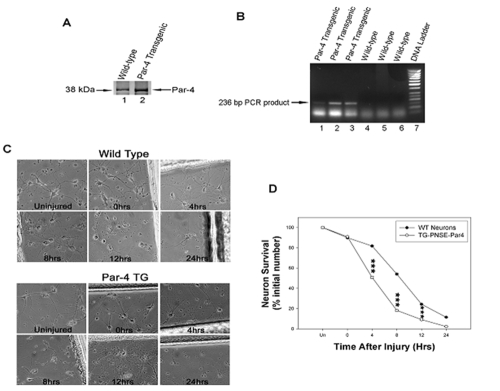
Traumatic brain injury (TBI) is a significant clinical problem, yet few effective strategies for treating it have emerged. People that sustain and survive a TBI are left with significant cognitive, behavioral, and communicative disabilities. Apoptotic neuronal death occurs following TBI. Prostate apoptosis response-4 (Par-4) is a death domain-containing protein initially characterized as a critical regulator of apoptosis in prostate cancer cells. We have recently generated and characterized Par-4 transgenic mice in which the expression of the par-4 transgene was limited to cells of neuronal lineage. We now provide evidence that, in cortical neurons from these mice, Par-4 drastically increases apoptotic neuronal death in both in vitro and in vivo models of TBI. In vitro experiments were performed in 7-day-old primary cultures of cortical neurons using a previously published, scratch-induced mechanical trauma model. Neurons that overexpress Par-4 showed not only a significant decrease in overall neuron survival after TBI compared to wild-type cells, but also exhibited a sharper decrease in mitochondrial transmembrane potential, a higher degree of free radical accumulation, and earlier activation of caspase-3 than wild-type cells did. In vivo experiments were performed utilizing a weight drop TBI model. A significantly increased volume of cortical injury and exacerbated activation of caspase-3 were observed in Par-4 transgenic mice when compared to those in wild-type mice. These data suggests that aberrant Par-4 expression exacerbates neuronal cell death following TBI by altering mitochondrial function, enhancing oxidative damage, and execution of apoptosis via caspase activation.

Keywords: Traumatic brain injury; apoptosis; cell culture; cerebral cortex; prostate apoptosis response-4.


Figures

Figure 1
Figure 1
Neurons that overexpress Par-4 show an exacerbated rate of cell death following the mechanical insult compared to wild-type neurons. A. Representative Western blotting analysis showing high level expression of Par-4 protein in cortical neurons from transgenic mice. Primary cultures of cortical neurons were established and western blot analyses were performed in 7-day old cultures using a monoclonal antibody raised against a recombinant protein corresponding to amino acids 1-334 representing full length PAR4 of rat origin (Santa Cruz Biotechnology, Inc.). Relatively high levels of Par-4 protein were detected in neurons from Par-4 transgenic mice (lane 2), while only minimal levels of endogenous Par-4 were observed in normal neurons from wild-type mice (lane 1). B. Genotyping of Par-4 transgenic mice using a PCR-based protocol. We have developed a quick PCR-based assay of the DNA from tail biopsies to amplify a 236-bp simian virus 40 fragment from pNSE-Par4 vector, which is detectable only in mice transgenic for Par-4 (lanes 1-3), but not in wild-type mice (lanes 4-6). C. Phase contrast images of cortical neurons from wild-type and Par-4 transgenic mice before and after injury. Overall neuron survival decreases at each time point. D. Graph shows percentage of surviving cells at each time point as indicated: uninjured (Un), immediately after injury (0), 4, 8, 12 and 24 hrs after injury. A higher percentage of cell death was observed in Par-4 transgenic cortical neurons compared to wild-type neurons. Values are the means and standard derivations of determinations from eight separate cultures (20-30 neurons analyzed per culture). ***p<0.001 compared with corresponding values in cells from wild-type mice. ANOVA with Scheffe's post-hoc tests.
Figure 2
Figure 2
Neurons that overexpress Par-4 exhibit enhanced mitochondrial dysfunction after mechanical injury compared to wild-type neurons. A. Rhd123 fluorescence (a measure of mitochondrial transmembrane potential) was measured before injury and at specific time intervals in both wild-type (top left) and Par-4 transgenic neurons (bottom left). Fluorescence intensity diminishes after injury, indicating the mitochondrial membrane potential has been compromised. B. Graph shows the mean average pixel intensity at each time point before and after injury. Par-4 transgenic neurons have significantly lower fluorescence intensity, beginning immediately after mechanical injury, when compared to wild-type neurons. Values are the means and standard derivations of determinations from eight separate cultures (20-30 neurons analyzed per culture). ***p<0.001 compared with corresponding values in cells from wild-type mice. ANOVA with Scheffe's post-hoc tests.
Figure 3
Figure 3
Cortical neurons overexpressing Par-4 show an increased accumulation of superoxide and hydrogen peroxide following mechanical injury compared to wild-type neurons. A. HE fluorescence (a measure of superoxide levels) and C. DCF fluorescence (a measure of hydrogen peroxide levels) were measured before injury and 12 hrs post injury in both wild-type (top) and Par-4 transgenic neurons (bottom). Fluorescence intensity of HE and DCF increases 12 hrs post injury in wild-type and Par-4 transgenic cortical neurons. B. Bar graph shows a higher accumulation of superoxide in neurons that overexpress Par-4 compared to wild-type neurons. D. Similar results are observed for the production of hydrogen peroxide in neurons overexpressing Par-4 when compared to wild-type neurons. Values are the means and standard derivations of determinations from eight separate cultures (20-30 neurons analyzed per culture). ***p<0.001 compared with corresponding values in cells from wild-type mice. ANOVA with Scheffe's post-hoc tests.
Figure 4
Figure 4
Par-4 overexpression in cortical neurons facilitates an earlier activation of caspase-3, contributing to a quicker progression through cell death. A. Levels of DEVD fluorescence (a measure of caspase-3 activation) were measured before injury and at specific time points after injury. Caspase-3 becomes activated after mechanical injury in both wild-type (top left) and Par4 transgenic neurons (bottom left). B. Quantification of DEVD fluorescence shows a higher degree of caspase-3 activation at each time point in Par-4 transgenic neurons compared to that in wild-type neurons, except at 24 hrs, when essentially all neurons overexpressing Par-4 have perished. Values are the means and standard derivations of determinations from eight separate cultures (20-30 neurons analyzed per culture). ***p<0.001. ANOVA with Scheffe's post-hoc tests.
Figure 5
Figure 5
Traumatic brain injury is exacerbated in Par-4 transgenic mice compared to wild-type mice. A. Coronal sections through the damaged cerebral cortex of adult mice were stained with cresyl violet. Nissl staining was measured in sham-operated mice and at 8, 24, and 48 hr intervals after cortical impact. Cortical damage is observed in the ipsilateral cortex (Ips), while the contralateral cortex (Ctl) is unaffected in wild-type (top left) and Par-4 transgenic mice (bottom left). B. Graph shows the percentage of cortical lesion volume in ipsilateral (Ips) and contralateral (Ctl) sides of the cortical impact in both wild-type and Par-4 transgenic mice. A significantly larger cavity was produced in the ipsilateral cortex (Ips) by 8 hrs and more prominently by 24 and 48 hrs after cortical impact in mice that overexpress Par-4 when compared to wild-type mouse brains. The damaged portion of the cortex was characterized by faint or lack of staining and was clearly delineated from healthy brain tissue. The amount of cortical damage is expressed as a percentage of total cortical volume, calculated using the Cavalieri principle (see Materials and Methods). Values are the means and standard derivation of determinations made from coronal brain sections through the area of impact and the contralateral side of impact. *p<.0.05, ***p<0.001 compared with corresponding values in sections from wild-type mice. ANOVA with Scheffe's post-hoc tests.
Figure 6
Figure 6
Caspase-3 activation is significantly greater in transgenic mice that overexpress Par-4 compared to wild-type mice. A. Coronal sections were immunostained with the mouse caspase-3 primary antibody, which only recognizes and labels the cleaved, activated form of caspase-3. Caspase-3 activation was measured in sham-operated mice and at 8, 12, and 24 hr intervals after weight drop. Caspase-3 activation increases at each time point in the ipsilateral cortex (Ips), while activation in the contralateral cortex (Ctl) remains unchanged in both wild-type (top left) and Par-4 transgenic mice (bottom left). B. Graph shows a higher degree of caspase-3 activation in Par-4 transgenic mice following cortical impact in the ipsilateral cortex (Ips), but not in the contralateral cortex (Ctl). Values are the means and standard derivations of determinations made from coronal brain sections through the area of impact and adjacent to the area of impact. ***p<0.001 compared with corresponding values in cells from wild-type mice. ANOVA with Scheffe's post-hoc tests.

Similar articles

Cited by

References

    1. DeFord SM, Wilson MS, Rice AC, Clausen T, Rice LK, Barabnova A, Bullock R, Hamm RJ. Repeated mild brain injuries result in cognitive impairment in B6C3F1 mice. J Neurotrauma. 2002;19:427–438. - PubMed
    1. Tweedie D, Milman A, Holloway HW, Li Y, Harvey BK, Shen H, Pistell PJ, Lahiri DK, Hoffer BJ, Wang Y, Pick CG, Greig NH. Apoptotic and behavioral sequelae of mild brain trauma in mice. J Neurosci Res. 2007;85:805–815. - PubMed
    1. Vink R, Nimmo AJ, Cernak I. An overview of new and novel pharmacotherapies for use in traumatic brain injury. Clin Exp Pharmacol Physiol. 2001;28:919–921. - PubMed
    1. Hatton J. Pharmacological treatment of traumatic brain injury: a review of agents in development. CNS Drugs. 2001;15:553–581. - PubMed
    1. Wong J, Hoe NW, Zhiwei F, Ng I. Apoptosis and traumatic brain injury. Neurocrit Care. 2005;3:177–182. - PubMed

LinkOut - more resources