. 2008 Jan 1;1(3):198-216.

Epstein-Barr virus and gastric carcinoma--viral carcinogenesis through epigenetic mechanisms

Affiliations

Epstein-Barr virus and gastric carcinoma--viral carcinogenesis through epigenetic mechanisms

Hiroshi Uozaki et al. Int J Clin Exp Pathol. .

Abstract

Epstein-Barr virus (EBV)-associated gastric carcinoma (GC) is the monoclonal growth of EBV-infected epithelial cells, and the entity was recognized only recently. EBV-associated GC is distributed worldwide and more than 90,000 patients are estimated to develop GC annually in association with EBV (10% of total GC). EBV-associated GC occurs in two forms in terms of the histological features, i.e., lymphoepithelioma-like GC and ordinary type of GC. Both share characteristic clinicopathological features, such as the preferential occurrence as multiple cancer and remnant stomach cancer. While the expression of EBV-latent genes is restricted to several in the infected cells (Latency I), EBV-associated GC shows gastric cell phenotype, resistance to apoptosis, and the production of immunomodulator molecules. Recently, global and non-random CpG island methylation of the promoter region of many cancer-related genes has been demonstrated with their decreased expression, such as p16 INK4A, p73 and E-cadherin. This abnormality is accompanied by methylation of the EBV genome itself, suggesting a process of virus-driven hypermethylation in the development of neoplastic cells. Further studies are necessary to determine the precise sequence of EBV infection, methylation, transformation and selection of the predominant clone within the stomach mucosa. Future studies are also desirable for the target and strategy of therapy, such as initiating viral replication or reversing the DNA methylation of cellular genes.

Keywords: DNA methylation; Epstein-Barr virus; chronic inflammation; gastric cancer; histology; viral oncogenesis.


Figures

Figure 1
Figure 1
Histology of an EBV-associated GC. This tumor is similar to lymphoepithelial carcinoma. Lymphocytes infiltrate massively among cancer cells (A, H&E stain). Lymphocytes are mainly CD8-positive T-cells. All tumor cells are positive for EBNA, whereas infiltrating lymphocytes are negative (B, EBER in situ hybridization).
Figure 2
Figure 2
Histology of an ordinary GC with EBV infection. About 5–10% of ordinary GCs are infected with EBV. EBV-associated GC has more lymphocyte infiltration than EBV-negative GC; HE sections (A for an example) give no suggestion of EBV infection. EBER in situ hybridization reveals EBV infection (B).
Figure 3
Figure 3
Macroscopic appearance of EBV-associated GC. EBV-associated GC often appears in the upper part of the stomach. This case is in the advanced stage. It is ulcerated without definite limits.
Figure 4
Figure 4
Features of non-neoplastic mucosa around GCs. EBV-associated GC is accompanied with atrophic gastritis and massive lymphocyte infiltration compared to both EBV-negative GC, diffuse type and intestinal type. H. pylori infection rate was not related to EBV infection.
Figure 5
Figure 5
Linear and circular episomal forms of EBV. EBV takes 2 forms, linear and circular. EBV replicates itself in the linear form, whereas EBV is in the state of latent infection. EBV binds at the terminal repeat (TR) and changes itself into the episomal circular form. Southern blotting after BamHI digestion with probes to TR reveals the different forms of EBV. The detected bands are short when EBV takes the linear form (a). When various clones of EBV infect the tumor, smearing signals are found (b). The band is single when monoclonal infection of EBV is established in the tumor (c).
Figure 6
Figure 6
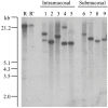
EBV clonality assay by Southern blot. DNA is cut with BamHI restriction enzyme, electrophoresed, and hybridized with an EBV-TR-specific probe. Five cases of intramucosal EBV-associated GCs and 4 cases of submucosal GC. Among intramucosal GC, 2 showed bi-clonal EBV infection and 3 showed monoclonal infection. All submucosal tumors showed monoclonal infection. EBV infection occurred at the initial or very early stage of carcinoma development.
Figure 7
Figure 7
Methylation rates of each gene examined by MSP. Seventeen cases of EBV-associated GC and 45 cases of EBV-negative GC were examined. EBV-associated GC showed a higher methylation rate of p14ARF, p15, p16INK4A, TIMP3, E-cad, DAPK, GSTP1 than EBV-negative GC.
Figure 8
Figure 8
Hypothesis of gastric carcinogenesis and maintenance by EBV. In healthy individuals, EBV infection of gastric epithelial cells is a very rare phenomenon (a). The entry point remains unclear. Even if EBV infected the gastric epithelium, usually EBV is cytotoxic and cannot persistently infect the gastric epithelium. Once persistent infection is established by an unknown trigger, the gastric epithelial cell methylates the EBV genome as a mechanism of host protection (b). The mechanism of DNA methylation works on the EBV genome, but also on the host genome in part. Some cells are methylated at the promoter region of tumor suppressor genes, and such cells dominantly proliferate (c, cancer initiation and progression). EBV infects tumor cells latently after EBV-associated GC is established. Latent genes of EBV maintain GC (d) through an anti-apoptotic effect or stimulation on growth factors. Some such effect makes the characteristic features of EBV-associated GC, such as a lace pattern, massive T cell infiltration, and expansive growth (e).

Similar articles

Cited by

References

    1. Epstein MA. Reflections on Epstein-Barr virus: some recently resolved old uncertainties. J Infect. 2001;43:111–115. - PubMed
    1. Burke AP, Yen TS, Shekitka KM, Sobin LH. Lymphoepithelial carcinoma of the stomach with Epstein-Barr virus demonstrated by polymerase chain reaction. Mod Pathol. 1990;3:377–380. - PubMed
    1. Min KW, Holmquist S, Peiper SC, O'Leary TJ. Poorly differentiated adenocarcinoma with lymphoid stroma (lymphoepithelioma-like carcinomas) of the stomach. Report of three cases with Epstein-Barr virus genome demonstrated by the polymerase chain reaction. Am J Clin Pathol. 1991;96:219–227. - PubMed
    1. Shibata D, Tokunaga M, Uemura Y, Sato E, Tanaka S, Weiss LM. Association of Epstein-Barr virus with undifferentiated gastric carcinomas with intense lymphoid infiltration. Lymphoepithelioma-like carcinoma. Am J Pathol. 1991;139:469–474. - PMC - PubMed
    1. Tokunaga M, Uemura Y, Tokudome T, Ishidate T, Masuda H, Okazaki E, Kaneko K, Naoe S, Ito M, Okamura A, et al. Epstein-Barr virus related gastric cancer in Japan: a molecular patho-epidemiological study. Acta Pathol Jpn. 1993;43:574–581. - PubMed

LinkOut - more resources