Review
. 2008 Nov;23(11):1399-407.
doi: 10.14670/HH-23.1399.

CXC chemokines and their receptors: a case for a significant biological role in cutaneous wound healing

Affiliations
Review

CXC chemokines and their receptors: a case for a significant biological role in cutaneous wound healing

Snjezana Zaja-Milatovic et al. Histol Histopathol. 2008 Nov.

Abstract

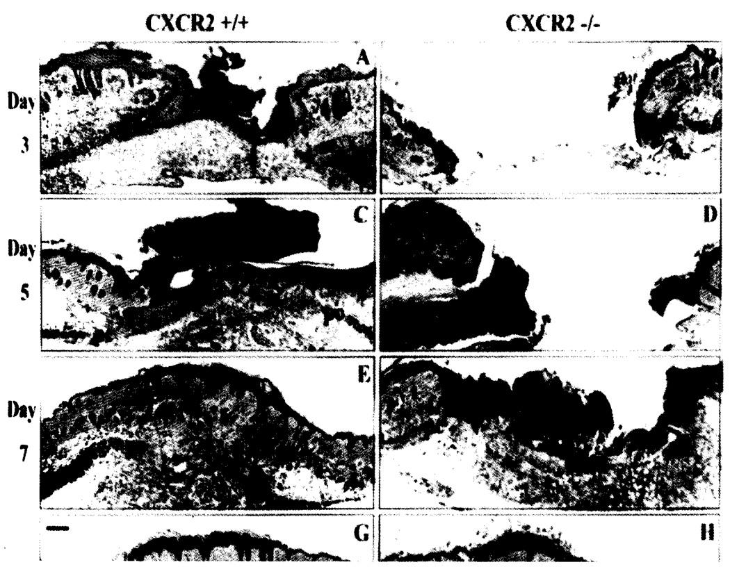
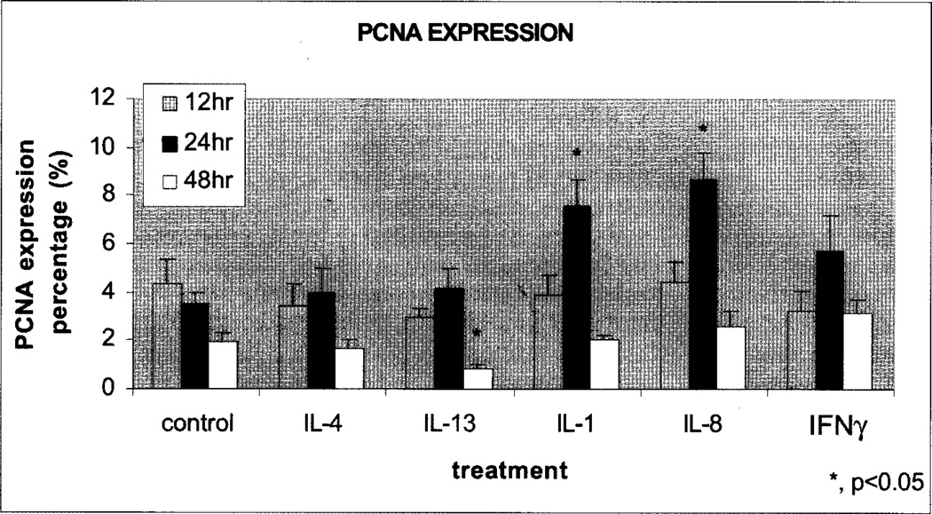
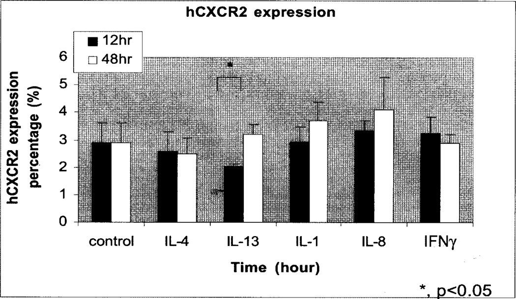
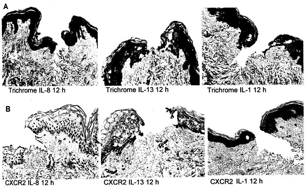
Wound healing requires a complex series of reactions and interactions among cells and their mediators, resulting in an overlapping series of events including coagulation, inflammation, epithelialization, formation of granulation tissue, matrix and scar formation. Cytokines and chemokines promote inflammation, angiogenesis, facilitate the passage of leukocytes from circulation into the tissue, and contribute to the regulation of epithelialization. They integrate inflammatory events and reparative processes that are important for modulating wound healing. Thus both cytokines and chemokines are important targets for therapeutic intervention. The chemokine-mediated regulation of angiogenesis is highly sophisticated, fine tuned, and involves pro-angiogenic chemokines, including CXCL1-3, 5-8 and their receptors, CXCR1 and CXCR2. CXCL1 and CXCR2 are expressed in normal human epidermis and are further induced during the wound healing process of human burn wounds, especially during the inflammatory, epithelialization and angiogenic processes. Human skin explant studies also show CXCR2 is expressed in wounded keratinocytes and Th/1/Th2 cytokine modulation of CXCR2 expression correlates with proliferation of epidermal keratinocytes. Murine excision wound healing, chemical burn wounds and skin organ culture systems are valuable models for examining the role of inflammatory cytokines and chemokines in wound healing.


Figures

Fig. 1
Fig. 1
Delayed epithelial resurfacing and wound closure in CXCR2−/− mice. Representative micrographs from trichome-stained sections in parts A-H show responses to excisional injury on days 3, 5, 7 and 10 post-injury. Parts A, C, E and G depict the normal sequence of repair in +/+ (wild-type) mice. Epithelialization underneath the vivid red scab is nearly complete by day 3 with a hypertrophic epidermis by day 5. Robust cellularity in the wound bed, increasing collagen deposition, and wound contracture are evident at days 5, 7 and 10. By contrast, wounds from CXCR2 knockout mice shown in B, D, F, and H reveal impaired reparative responses: Resurfacing across the wound bed remains incomplete until days 7–8. Severely inhibited cellularity of the granulation tissue (gt) is noted at days 3–5 and delays in wound contracture are evident at days 3, 5 and 7. Immature granulation tissue is visible at day 7 followed by eventual wound closure and complete resurfacing by day 10. All wounds eventually show complete epidermal and dermal healing with no scarring. After resurfacing is achieved, sites of excisional wounding can only be detected by a discontinuity in the underlying panniculus-camosus layer (pc). Scale bars: 100 µm. (Reprinted with permission from Devalaraja et al., J. Invest. Dermatol. (2000) 115, 234–244.)
Fig. 2
Fig. 2
Evaluation of the proliferative cellular nuclear antigen (PCNA) in human skin explant cultures treated with cytokines. Paraformaldehyde fixed, paraffin embedded human skin tissue sections were immunostained for proliferating cell nuclear antigen (PCNA). PCNA immunostaining showed a significant decrease (p<0.05) in the number of proliferating cells in the skin tissue explants treated with IL-13 after 48 hours of cytokine treatment. Treatments with IL-1 and IL-8 showed significant increase (p<0.05) in the number of proliferative cells (p<0.05) after 24 hours as compared to untreated control.
Fig. 3
Fig. 3
Expression of hCXCR2 in the epidermis of human skin explant cultures treated with cytokines. Paraformaldehyde fixed, paraffin embedded human skin tissue sections were immunostained for hCXCR2. IL-13 showed a significant decrease (p<0.05) in the number of CXCR2 positive cells after 12 hours of treatment as compared to untreated control.
Fig. 4
Fig. 4
A. Histological analysis of wounded human skin explants treated with IL-8, IL-13. or IL-1. Trichrome stained human skin tissue samples showed new collagen and immature epidermal cells at 12 hours after injury. Pictures were taken at 10× magnification. B. Immunohistochemical analysis of CXCR2 expression in IL-1, IL-13, and IL-8 treated human skin tissue explants. Analysis showed that IL-13 significantly decreased the number of CXCR2 positive cells after 12 hours of treatment as compared as IL-1 and IL-8.

References

    1. Akasaka Y, Ono I, Yamashita T, Jimbow K, Ishii T. Basic fibroblast growth factor promotes apoptosis and suppresses granulation tissue formation in acute incisional wounds. J. Pathol. 2004;203:710–720. - PubMed
    1. Anitua E, Andia I, Ardanza B, Nurden P, Nurden AT. Autologous platelets as a source of proteins for healing and tissue regeneration. Thromb. Haemost. 2004;91:4–15. - PubMed
    1. Balkwill F, Manotovani A. Inflammation and cancer: back to Virchow? Lancet. 2001;357:539–545. - PubMed
    1. Banchereau J. Converging and diverging properties of human interleukin-4 and interleukin-10. Boehring Inst. Mitt. 1995:58–77. - PubMed
    1. Bonecchi R, Facchetti F, Dusi S, Luini W, Lissandrini D, Simmelink M, Locati M, Bernasconi S, Allavena P, Brandt E, Rossi F, Mantovani A, Sozzani S. Induction of functional IL-8 Receptors by IL-4 and IL-13 in Human Monocytes. J. Immunol. 2000;164:3862–3869. - PubMed

Publication types