. 2008 Oct 7;47(40):10694-704.
doi: 10.1021/bi800533t. Epub 2008 Sep 12.

Probing the pH-dependent prepore to pore transition of Bacillus anthracis protective antigen with differential oxidative protein footprinting

Affiliations

Probing the pH-dependent prepore to pore transition of Bacillus anthracis protective antigen with differential oxidative protein footprinting

James G Smedley 3rd et al. Biochemistry. .

Abstract

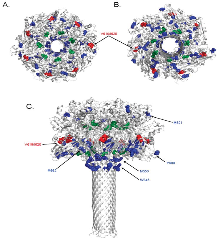
The protective antigen (PA) component of the anthrax toxin (ATx) plays an essential role in the pathogenesis of the bioterrorism bacterium Bacillus anthracis. After oligomerization on the cell surface and docking of lethal factor and/or edema factor, PA is internalized and undergoes a conformational change when exposed to the low pH of the endosome to form a membrane-penetrating pore. While the structure of the PA prepore has been determined, precise structural information regarding the pore state remains lacking. Oxidative protein footprinting (OPF) can provide dynamic structural information about a protein complex through analysis of amino acid oxidation both before and after a conformational change. In this study, PA at pH 7.5 and 5.5 was exposed to hydroxyl radicals generated by ionizing radiation. Mass spectrometry was then used to both identify and quantitate the extent of oxidation of differentially modified residues. Several residues were found to be more readily oxidized at pH 7.5, most of which clustered toward the bottom plane of the prepore heptamer. Two amino acids had greater oxidation rates at pH 5.5, both found on the outer periphery of the prepore. When the OPF results were mapped to a current computational model of the pore, the accessibilities of some residues were consistent with their modeled positions in the pore (i.e., Y688 and V619/I620), while data for other residues (W346 and M350) appeared to conflict with the model. The results from this study illustrate the utility of OPF in generating empirical structural information for yet undetermined structures and offering opportunities for refinement for models thereof.


Figures

Figure 1
Figure 1
Structural map of PA. (A) Primary amino acid structure of PA highlighting domains 1 (pink), 2 (green), 3 (yellow), and 4 (blue). Proteolytic fragment PA20 consists of amino acids 1–167 and is italicized. Residues found to be oxidized in this study are in red and bold type, and tryptic fragments containing those residues are shown in brackets. Amino acid coverage by MS/MS analysis is shown with a dashed underline. (B) The 2.1 Å three-dimensional crystal structure of PA [PDB entry 1ACC (18)] with each of its four domains colored as in panel A. The disordered loop that forms part of the transmembrane stem domain (residues 302–325) is shown as a dashed line.
Figure 2
Figure 2
Quantitative analysis of oxidation from ESI-MS. The amount of peptide oxidation was calculated for each PA63 amino acid found modified by MS/MS analysis. For each oxidized peptide under each experimental condition, the signal abundance of both unoxidized and oxidized ions was recorded, each in triplicate. (A) Data are expressed as the percent total oxidized peptide [oxidized/ (oxidized + unoxidized) × 100], and error bars represent the calculated standard deviation. (B) The increase in the level of oxidation for indicated PA63 residues was calculated by dividing the percent total oxidized peptide after irradiation by that of control samples. Shown are representative data for each of the amino acids in this study for which quantitation of oxidation was carried out.
Figure 3
Figure 3
Mapping oxidized residues on the PA63 prepore. Amino acids found to be oxidized in this study are shown in colored spheres on a ribbon rendering of the 3.6 Å crystal structure of the PA63 prepore heptamer [PDB entry 1TZO (17)]. Residues observed to be oxidized more readily at pH 7.5 are colored blue, residues found to oxidized more readily at pH 5.5 red, and residues which demonstrated no change in oxidation between the two pH values green. Depicted in this figure are (A) a side view (with the “bottom plane” identified with a dashed line), (B) a zoomed view of the lower bottom plane, (C) a top-down view, and (D) a bottom-up view.
Figure 4
Figure 4
Anisotropic temperature factors of the PA63 prepore heptamer. The atoms of the PA63 prepore crystal structure [PDB entry 1TZO (17)] are colored on the basis of their anisotropic temperature factor. The spectrum from blue to red represents atoms with low to high b-factor values, respectively, relative to all atoms in the PA63 prepore structure. Sites identified to be oxidized in this study are represented as spheres. Depicted in this figure are side (A), bottom-up (B), and top-down (C) views of the prepore heptamer.
Figure 5
Figure 5
Mapping oxidized residues on the PA63 pore model. Amino acids found to be oxidized in this study are shown as colored spheres on a ribbon rendering of three-dimensional model of the PA63 pore heptamer [PDB entry 1V36 (47)]. Residues observed to be oxidized more readily at pH 7.5 are colored blue, residues found to oxidized more readily at pH 5.5 red, and residues which demonstrated no change in oxidation between the two pH values green. Depicted in this figure are top-down (A), bottom-up (B), and side (C) views of the pore heptamer.

Similar articles

Cited by

References

    1. Mogridge J, Cunningham K, Lacy DB, Mourez M, Collier RJ. The lethal and edema factors of anthrax toxin bind only to oligomeric forms of the protective antigen. Proc Natl Acad Sci USA. 2002;99:7045–7048. - PMC - PubMed
    1. Bradley KA, Mogridge J, Mourez M, Collier RJ, Young JA. Identification of the cellular receptor for anthrax toxin. Nature. 2001;414:225–229. - PubMed
    1. Scobie HM, Rainey GJ, Bradley KA, Young JA. Human capillary morphogenesis protein 2 functions as an anthrax toxin receptor. Proc Natl Acad Sci USA. 2003;100:5170–5174. - PMC - PubMed
    1. Klimpel KR, Molloy SS, Thomas G, Leppla SH. Anthrax toxin protective antigen is activated by a cell surface protease with the sequence specificity and catalytic properties of furin. Proc Natl Acad Sci USA. 1992;89:10277–10281. - PMC - PubMed
    1. Molloy SS, Bresnahan PA, Leppla SH, Klimpel KR, Thomas G. Human furin is a calcium-dependent serine endoprotease that recognizes the sequence Arg-X-X-Arg and efficiently cleaves anthrax toxin protective antigen. J Biol Chem. 1992;267:16396–16402. - PubMed

Publication types

MeSH terms