. 2008 Nov;9(11):1894-904.
doi: 10.1111/j.1600-0854.2008.00810.x. Epub 2008 Aug 9.

Acute perturbations in Golgi organization impact de novo sphingomyelin synthesis

Affiliations

Acute perturbations in Golgi organization impact de novo sphingomyelin synthesis

Suchismita Chandran et al. Traffic. 2008 Nov.

Abstract

The mammalian Golgi apparatus is composed of multiple stacks of cisternal membranes organized laterally into a ribbon-like structure, with close apposition of trans Golgi regions with specialized endoplasmic reticulum (ER) membranes. These contacts may be the site of ceramide transfer from its site of synthesis (ER) to sphingomyelin (SM) synthase through ceramide transfer protein (CERT). CERT extracts ceramide from the ER and transfers it to Golgi membranes but the role of overall Golgi structure in this process is unknown. We show here that localization of CERT in puncta around the Golgi complex requires both ER- and Golgi-binding domains of CERT. To examine how Golgi structure contributes to SM synthesis, we treated cells with Golgi-perturbing drugs and measured newly synthesized SM. Interestingly, disruption of Golgi morphology with nocodazole, but not ilimaquinone inhibited SM synthesis. Decreased localization of CERT with a Golgi marker correlated with decreased SM synthesis. We propose that some Golgi structural perturbations interfere with efficient ceramide trafficking through CERT, and thus SM synthesis. The organization of the mammalian Golgi ribbon together with CERT may promote specific ER-Golgi interactions for efficient delivery of ceramide for SM synthesis.


Figures

Figure 1
Figure 1
Distribution of endogenous CERT in HeLa cells. HeLa cells expressing galT-DsRed (Golgi marker) and CFP-ER (ER marker) were treated with DMSO (vehicle control) for 1 h at 37°C in presence of 10 μg/ml cycloheximide to mimic lipid labeling experiments. Cells were stained with commercially available chicken anti-CERT IgY (IgG) and Cy5 conjugated anti-chicken IgY (IgG) antibodies. The cells were imaged by confocal microscopy and Volocity software was used to create a single image from the confocal sections. Arrows indicate CERT-ER marker colocalization. Bars, 10 μm.
Figure 2
Figure 2
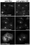
ER and Golgi binding domains of CERT are required for its steady state localization. HeLa cells were transfected with plasmids encoding Myc-tagged versions of wild type CERT, CERT-FFATmut, or CERT-PHmut, as indicated. Cells were stained with affinity purified rabbit anti-golgin-160 antibodies (right panels) as a marker for the Golgi apparatus and CERT was identified by mouse anti-Myc antibodies (left panels). Secondary antibodies were Texas red conjugated goat anti-rabbit (IgG) and Alexa-488 conjugated donkey anti-mouse (IgG). Bars, 10 μm.
Figure 3
Figure 3
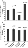
Newly synthesized sphingomyelin and ceramide levels in cells treated with Golgi perturbing drugs. HeLa cells were labeled with 3H-serine and incubated with ilimaquinone (2.5 μg/ml), nocodazole (2.5 μg/ml) or brefeldin-A (1 μg/ml) for 1 h at 37°C in presence of 10 μg/ml cycloheximide. DMSO or ethanol were used as vehicle controls for the drugs. Lipids were extracted and analyzed as described in Materials and Methods. The levels of newly synthesized sphingomyelin (A) and ceramide (B) were normalized to the total amount of protein and are indicated as a percentage of the level in vehicle-treated control. The data represent 6 independent experiments for ilimaquinone and nocodazole treatment, and 5 independent experiments for brefeldin-A. Standard error of the mean was calculated for each treatment and is represented on the graph. Student’s T-test values (p), where the data from drug treatments were compared to their respective controls are indicated above each column.
Figure 4
Figure 4
Localization of CERT is altered in cells treated with Golgi perturbing drugs. HeLa cells expressing galT-DsRed (trans Golgi marker), CFP-ER (ER marker) and Myc-tagged CERT were treated with (A) DMSO (vehicle control), (B) ilimaquinone (2.5 μg/ml) or (C) nocodazole (2.5 μg/ml) for 1 h at 37°C in presence of 10 μg/ml cycloheximide to mimic the lipid labeling experiments. After fixation and staining for CERT with anti-Myc and Cy5 conjugated anti-mouse (IgG), confocal microscopy was performed and 3-D images were created using Volocity software. Note that the ER appears fragmented by the fixation method used here. A higher magnification of the selected regions is shown in the lower panels for each of the treatments. The enlarged region represents several optical sections of the entire stack of sections that was collected. Sections 2–4 out of a total of 6 sections are shown in the enlargement in DMSO treated cells, sections 3–4 out of a total of 5 sections are shown in the enlargement in ilimaquinone-treated cells and sections 7–10 out of a total of 13 sections are shown in the enlargement in nocodazole-treated cells. Arrows in (B) indicate CERT-Golgi marker colocalization. Bars, 10 μm.
Figure 5
Figure 5
Decreased overlap of CERT with Golgi membranes in nocodazole-treated cells. Overlap of CERT with the Golgi (galT-DsRed) and ER (CFP-ER) markers was quantified for 9–10 individual cells for each treatment as described in Materials and Methods, and the overlap coefficient was calculated. The graph indicates average overlap coefficient values for each treatment after normalization to the DMSO control (which was set to 1.0). Open columns indicate overlap coefficient of CERT with the Golgi marker, and closed columns indicate overlap coefficient of CERT with the ER marker. Standard error of the mean was calculated for each treatment and is represented on the graph.
Figure 6
Figure 6
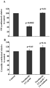
Newly synthesized SM levels recover after nocodazole washout. Cells treated with nocodazole (2.5 μg/ml) for 1 h were washed and cultured in the absence of drug for 2 h, and labeled with 3H-serine during the last 1 h. Lipids were extracted and analyzed as described in Materials and Methods. The levels of newly synthesized sphingomyelin (A) and ceramide (B) were normalized to the total amount of protein and are indicated as a percentage of the level in vehicle-treated control. The data represent 3 independent experiments. Standard error of the mean was calculated for each treatment and is represented on the graph. Student’s T-test values (p), where data from each treatment were compared to the vehicle-treated control are indicated above each column.
Figure 7
Figure 7
Nocodazole does not inhibit SM synthase activity, alter phosphorylation of CERT or eliminate CERT’s ability to associate with Golgi membranes. (A) Nocodazole does not inhibit SM synthase activity. Hela cell lysates were incubated with nocodazole (2.5 μg/ml) and 5 nmol NBD-C6-Cer for 30 min at 30°C. DMSO was used as the control for nocodazole. Fluorescent sphingomyelin was quantified as described in Materials and Methods. The data represent 4 independent experiments. Standard error of the mean was calculated for each treatment and is represented on the graph. Student’s T-test values (p), where the data from nocodazole-treated lysates were compared to control lysates are indicated above each column. (B) Nocodazole does not affect the phosphorylation status of CERT. HeLa cells were transfected with plasmids encoding Myc-tagged CERT for 24 h and treated with DMSO (vehicle control) or nocodazole (2.5 μg/ml) for 1 h at 37°C. Lysates were incubated in the absence or presence of lambda phosphatase, separated by SDS-PAGE and immunoblotted for CERT with anti-Myc antibody. (C) Nocodazole does not eliminate CERT’s ability to associate with Golgi membranes. HeLa cells were transfected with plasmids encoding the Golgi marker galT-DsRed and Myc-tagged wild type CERT (wt-CERT) or CERT-FFATmut as indicated. Cells were incubated with nocodazole (2.5 μg/ml) for 1 h at 37°C. CERT proteins were identified by indirect immunofluorescence microscopy using anti-Myc antibody and Alexa-488 conjugated donkey anti-mouse secondary antibody (IgG). Bars, 10 μm. The graph on the right represents the overlap coefficient of CERT with the Golgi marker for 8–22 individual cells for each treatment as described in Materials and Methods. Standard error of the mean was calculated for each treatment and is represented on the graph.

Similar articles

Cited by

References

    1. Farquhar MG, Palade GE. The Golgi apparatus: 100 years of progress and controversy. Trends Cell Biol. 1998;8(1):2–10. - PMC - PubMed
    1. Ohanian J, Ohanian V. Sphingolipids in mammalian cell signalling. Cell Mol Life Sci. 2001;58(14):2053–2068. - PMC - PubMed
    1. Cutler RG, Mattson MP. Sphingomyelin and ceramide as regulators of development and lifespan. Mech Ageing Dev. 2001;122(9):895–908. - PubMed
    1. Futerman AH, Riezman H. The ins and outs of sphingolipid synthesis. Trends Cell Biol. 2005;15(6):312–318. - PubMed
    1. Halter D, Neumann S, van Dijk SM, Wolthoorn J, de Maziere AM, Vieira OV, Mattjus P, Klumperman J, van Meer G, Sprong H. Pre- and post-Golgi translocation of glucosylceramide in glycosphingolipid synthesis. J Cell Biol. 2007;179(1):101–115. - PMC - PubMed

Publication types

Substances

LinkOut - more resources