

The complement cascade: Yin-Yang in neuroinflammation--neuro-protection and -degeneration
- PMID: 18786171
- PMCID: PMC4038542
- DOI: 10.1111/j.1471-4159.2008.05668.x
The complement cascade: Yin-Yang in neuroinflammation--neuro-protection and -degeneration
Abstract
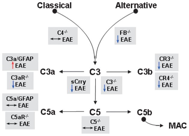
The complement cascade has long been recognized to play a key role in inflammatory and degenerative diseases. It is a 'double edged' sword as it is necessary to maintain health, yet can have adverse effects when unregulated, often exacerbating disease. The contrasting effects of complement, depending on whether in a setting of health or disease, is the price paid to achieve flexibility in scope and degree of a protective response for the host from infection and injury. Loss or even decreased efficiency of critical regulatory control mechanisms can result in aggravated inflammation and destruction of self-tissue. The role of the complement cascade is poorly understood in the nervous system and neurological disorders. Novel studies have demonstrated that the expression of complement proteins in brain varies in different cell types and the effects of complement activation in various disease settings appear to differ. Understanding the functioning of this cascade is essential, as it has therapeutic implications. In this review, we will attempt to provide insight into how this complex cascade functions and to identify potential strategic targets for therapeutic intervention in chronic diseases as well as acute injury in the CNS.
Figures


References
-
- Abbott NJ, Mendonca LL, Dolman DE. The blood–brain barrier in systemic lupus erythematosus. Lupus. 2003;12:908–915. - PubMed
-
- Afagh A, Cummings BJ, Cribbs DH, Cotman CW, Tenner AJ. Localization and cell association of C1q in Alzheimer’s disease brain. Exp Neurol. 1996;138:22–32. - PubMed
-
- Ahmed F, Brown KM, Stephan DA, Morrison JC, Johnson EC, Tomarev SI. Microarray analysis of changes in mRNA levels in the rat retina after experimental elevation of intraocular pressure. Invest Ophthalmol Vis Sci. 2004;45:1247–1258. - PubMed
-
- Alexander JJ, Quigg RJ. Systemic lupus erythematosus and the brain: what mice are telling us. Neurochem Int. 2007;50:5–11. - PubMed
-
- Alexander JJ, Bao L, Jacob A, Kraus DM, Holers VM, Quigg RJ. Administration of the soluble complement inhibitor, Crry-Ig, reduces inflammation and aquaporin 4 expression in lupus cerebritis. Biochim Biophys Acta. 2003;1639:169–176. - PubMed
Publication types
MeSH terms
Substances
Grants and funding
LinkOut - more resources
Full Text Sources
Other Literature Sources
Medical
