

Losses of expression of the antigens A, Lea and Lex and over-expression of Ley in carcinomas and HG-SIL of the uterine cervix
- PMID: 18786253
- PMCID: PMC2551588
- DOI: 10.1186/1746-1596-3-38
Losses of expression of the antigens A, Lea and Lex and over-expression of Ley in carcinomas and HG-SIL of the uterine cervix
Abstract
Background: The glycosylation of a great number of molecules, glyco-protein or glycolipids, has been of interest for decades.
Objective: To compare the expressive patterns of the isoantigenic determinants of histo-blood groups ABH and Lewis in squamous and simple epithelium and in precursors and cancers of the cervix.
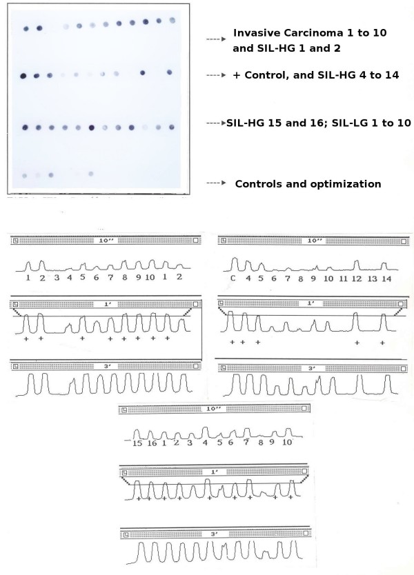
Methods: A total of 36 lesions and neoplasms (10 LG-SIL, 16 HG-SIL and 10 invasive carcinomas) have been studied with immunohistochemical techniques, using monoclonal antibodies (MoAb BG1 to BG8) for precursor chains, blood-group ABH and Lewis group Le(a), Le(b), Le(x), and Le(y), and four types of lectins. In addition, we have studied the expression of p53 protein and PCNA, establishing the rate of proliferation of each lesion. Using PCR techniques, we have also detected part of the intron of the E6 gene of HPV-16.
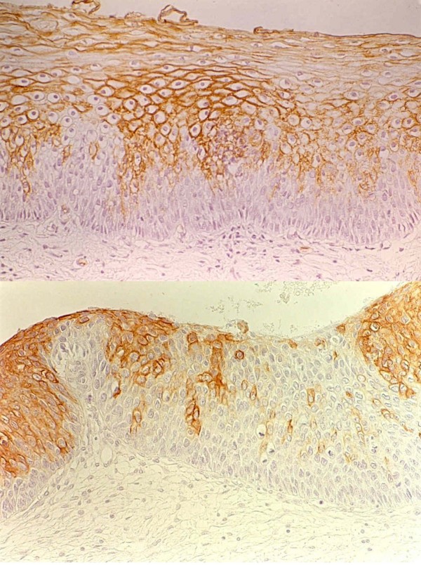
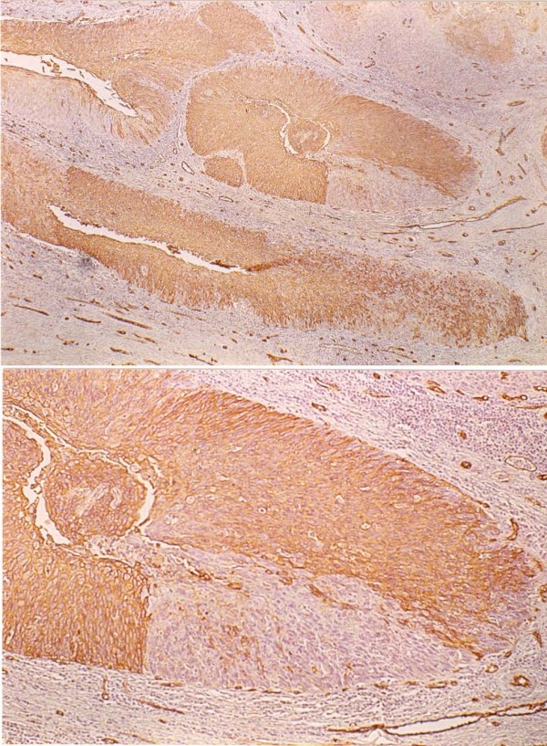
Results: In the invasive cervical carcinomas, we observed a loss of expression of the Le(x) antigen (p < 0.01). With regard to the progression of the different lesions studied, we found alterations in the patterns of expression of the antigens of the ABH and Lewis blood groups. There was a tendency towards a loss of expression and heterogeneous patterns in the more advanced lesions, as well as over-expression of the Le(y) antigens. With PCNA, we established a proliferative rate which tended to be greater in relation to the progression of the cervix neoplasms.
Conclusion: These results indicate that there is a relation between the losses of histo-blood groups and the progression of the squamous intraepithelial lesions.
Figures


Similar articles
-
ABH and related histo-blood group antigens in normal & malignant human endometrium in relation to genetic and hormonal factors.APMIS Suppl. 1997;69:1-33. APMIS Suppl. 1997. PMID: 9254838
-
Expression of blood group-related antigens ABH, Lewis A, Lewis B, Lewis X, Lewis Y, and CA 19-9 in pancreatic cancer cells in comparison with the patient's blood group type.Cancer Res. 1988 Oct 1;48(19):5422-6. Cancer Res. 1988. PMID: 3166398
-
Presence of two distinct acinar cell populations in human pancreas based on their antigenicity.Int J Pancreatol. 1986 Oct;1(3-4):213-25. doi: 10.1007/BF02795247. Int J Pancreatol. 1986. PMID: 3316426
-
Glycan structures and their recognition roles in the human blood group ABH/Ii, Lea, b, x, y and Sialyl Lea,x active cyst glycoproteins.Glycoconj J. 2019 Dec;36(6):495-507. doi: 10.1007/s10719-019-09887-x. Epub 2019 Nov 26. Glycoconj J. 2019. PMID: 31773366 Review.
-
ABH and related histo-blood group antigens; immunochemical differences in carrier isotypes and their distribution.Vox Sang. 1989;56(1):1-20. doi: 10.1111/j.1423-0410.1989.tb03040.x. Vox Sang. 1989. PMID: 2464874 Review.
Cited by
-
Differential fucosyltransferase IV expression in squamous carcinoma cells is regulated by promoter methylation.Cell Mol Biol Lett. 2012 Jun;17(2):206-16. doi: 10.2478/s11658-012-0003-x. Cell Mol Biol Lett. 2012. PMID: 22287018 Free PMC article.
-
Sialyl Lewis x expression in cervical scrapes of premalignant lesions.J Biosci. 2012 Dec;37(6):999-1004. doi: 10.1007/s12038-012-9261-z. J Biosci. 2012. PMID: 23151790
-
ABO/Rh Blood Group and Cervical Cancer Survival: Results from Our Own and Other Studies.J Cancer. 2024 Jul 9;15(15):4777-4788. doi: 10.7150/jca.95245. eCollection 2024. J Cancer. 2024. PMID: 39132152 Free PMC article.
-
Blood group antigens SLeX, SLeA, and LeY as prognostic markers in endometrial cancer.J Cancer Res Clin Oncol. 2022 Dec;148(12):3323-3335. doi: 10.1007/s00432-022-04098-8. Epub 2022 Jun 21. J Cancer Res Clin Oncol. 2022. PMID: 35729354 Free PMC article.
-
Glycomics of cervicovaginal fluid from women at risk of preterm birth reveals immuno-regulatory epitopes that are hallmarks of cancer and viral glycosylation.Sci Rep. 2024 Sep 6;14(1):20813. doi: 10.1038/s41598-024-71950-x. Sci Rep. 2024. PMID: 39242814 Free PMC article.
References
-
- Hakomori S-I. Preface to Carbohydrate Pathology. Acta Path Mic Sc. 1992;100:18–27. - PubMed
-
- Clausen H, Hakomori S. ABH and related histo-blood group antigens; inmunohistochemical differentiation in carrier isotypes and their distribution. Vox Sang. 1989;56:1–20. - PubMed
-
- Holgersson J, Breimmer ME, Samuelsson BE. Basic biochemistry of cell surface carbohydrates and aspects of the tissue distribution of histo-blood group ABH and related glycosphingolipids. APMIS Suppl. 1992;27:18–27. - PubMed
-
- Hakomori S-I. In: General concept of tumor-associated carbohydrate antigens: Their chemical, phisical and enzymatic basis. In: Gangliosides and Cancer. Oettgen HF, editor. V.C.H; 1989. pp. 58–68.
LinkOut - more resources
Full Text Sources
Research Materials
Miscellaneous
