. 2008 Sep 10;16(9):1399-406.
doi: 10.1016/j.str.2008.05.016.

Bacteriophage lambda stabilization by auxiliary protein gpD: timing, location, and mechanism of attachment determined by cryo-EM

Affiliations

Bacteriophage lambda stabilization by auxiliary protein gpD: timing, location, and mechanism of attachment determined by cryo-EM

Gabriel C Lander et al. Structure. .

Abstract

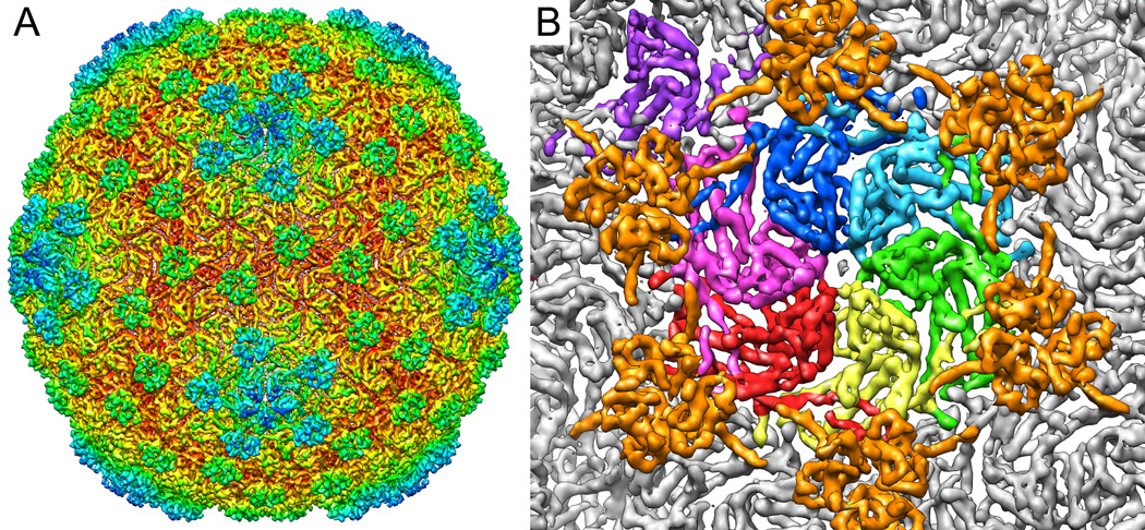
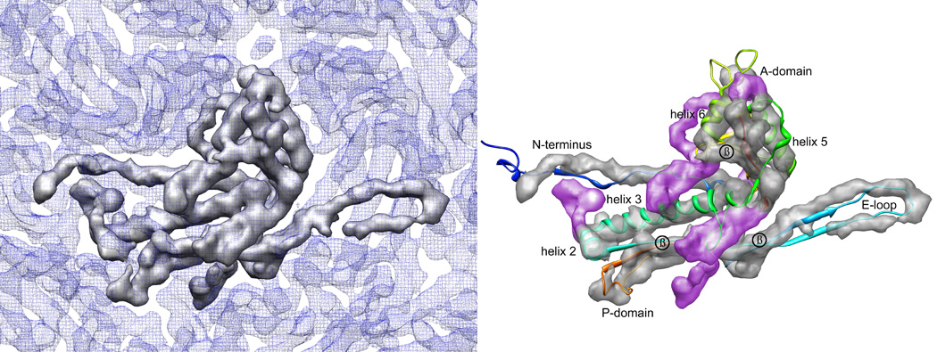
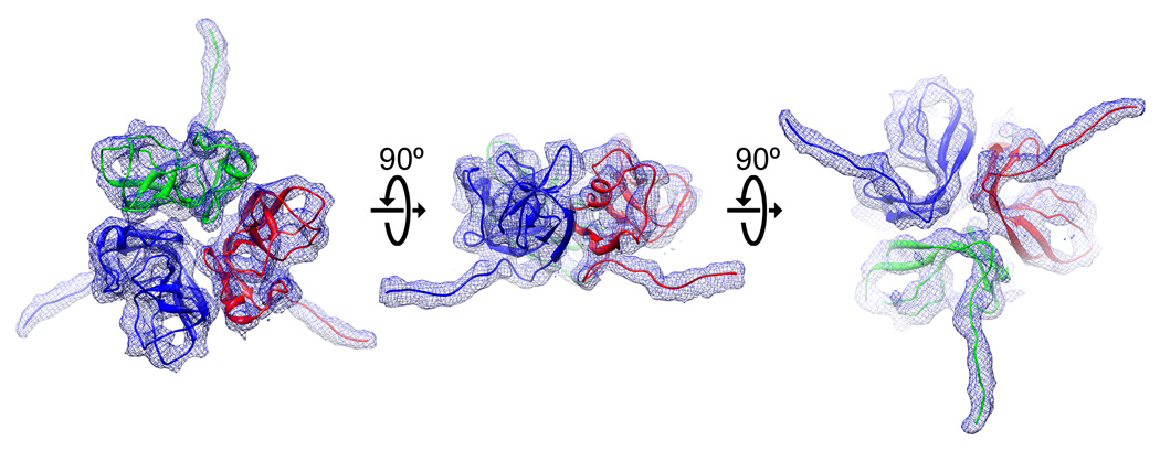
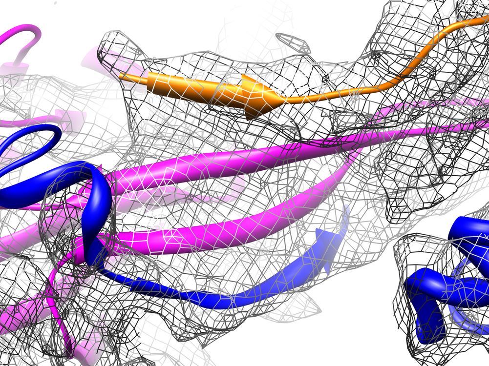
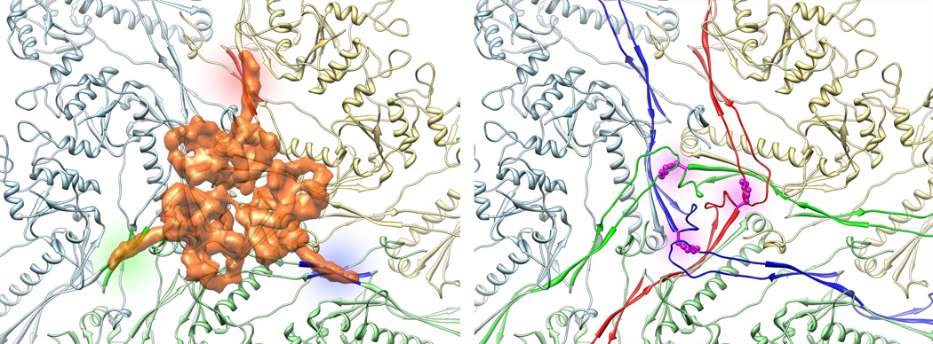
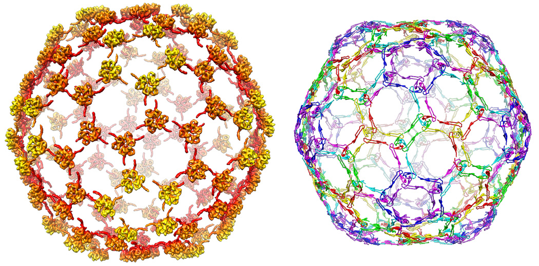
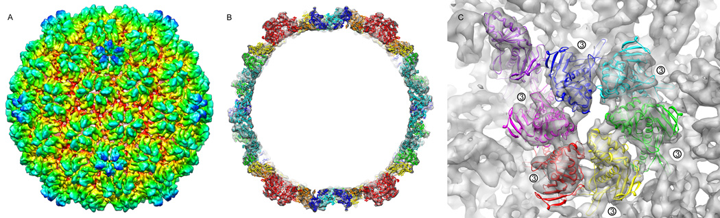
We report the cryo-EM structure of bacteriophage lambda and the mechanism for stabilizing the 20-A-thick capsid containing the dsDNA genome. The crystal structure of the HK97 bacteriophage capsid fits most of the T = 7 lambda particle density with only minor adjustment. A prominent surface feature at the 3-fold axes corresponds to the cementing protein gpD, which is necessary for stabilization of the capsid shell. Its position coincides with the location of the covalent cross-link formed in the docked HK97 crystal structure, suggesting an evolutionary replacement of this gene product in lambda by autocatalytic chemistry in HK97. The crystal structure of the trimeric gpD, in which the 14 N-terminal residues required for capsid binding are disordered, fits precisely into the corresponding EM density. The N-terminal residues of gpD are well ordered in the cryo-EM density, adding a strand to a beta-sheet formed by the capsid proteins and explaining the mechanism of particle stabilization.


Figures

Figure 1
Figure 1. Three-dimensional density of mature bacteriophage lambda reconstructed from CryoEM micrographs
A) The subnanometer-resolution map of bacteriophage lambda colored radially from the phage center (red to blue). The reconstruction shows the T=7 laevo symmetry of the capsid, and the decoration protein gpD as protruding densities at the quasi three and six fold vertices. B) A close up, segmented view of the seven subunits that make up the icosahedral asymmetric unit, colored by subunit. The pentameric subunit is seen in the upper left-hand corner in purple. Surrounding the asymmetric unit are six gpD molecules, colored orange.
Figure 2
Figure 2. Segmentation of one monomer of gpE
A) One capsid subunit is shown as a surface representation in grey, in the context of the surrounding density, in blue mesh. B) Rigid-body fitting of HK97 crystal structure into the lambda monomer density, demonstrating closely similar morphology. For this fitting, the A domain of HK97 (residues 242–332, 373–383) was separated from the rest of the crystal structure and fit independently, rotated 15° clockwise relative to HK97. Salient features observed in this map include homologous densities for helices 2, 3, 5, and 6, as well as putative beta-sheet densities, labeled in the figure. Additional density unaccounted for by the HK97 crystal structure (colored in purple) can be attributed to the additional 59 residues the lambda gpE contains in comparison to HK97.
Figure 3
Figure 3. Isolated density corresponding to gpD, with modeled N-terminal residues
Top, side, and bottom views of gpD are displayed with the 1.1 Å crystal structure (PDB id: 1C5E) fit into the reconstructed EM density, colored by subunit. Poly-alanine peptides of the fourteen disordered residues of the N-terminus were modeled into the density, which can be seen extending away from the main body of the gpD trimer. It is evident from these views why the N-terminus is disordered in the absence of the mature capsid substrate.
Figure 4
Figure 4. The stabilizing four-stranded beta sheet
During maturation of phage lambda, the E-loop of one gpE capsid protein (magenta) interacts with the N-terminus of a neighboring gpE subunit within the same capsomer (blue), forming a beta sheet similar to that seen in HK97. Although the individual polypeptide strands that make up the beta sheet are not distinguishable in the EM density, this density accommodates the HK97 three-stranded beta sheet. An additional strand is contributed by gpD (orange) as is binds to the capsid surface, shown by the contribution of an additional density above the three capsid strands.
Figure 5
Figure 5. Homologous stabilization method in lambda and HK97
A three-fold vertex of the HK97 crystal structure is shown as a ribbon, each capsomer colored differently (light blue, yellow, and green). On the left, density from the reconstruction corresponding to gpD has been overlaid semi-transparently as it is situated in lambda. Note that although lambda does not undergo covalent crosslink formation, gpD’s N-terminal arms form strong beta-sheet interactions with each of three capsomers, securing the capsid at the three-fold. Sites of interaction are colored in red, blue and green. The HK97 lysine-asparagine crosslink, colored in magenta on the right, secure the three capsomers covalently, such that a cementing protein was no longer necessary. The polypeptide arms involved in creation of the HK97 chainmail are colored in red, blue and green.
Figure 6
Figure 6. Comparison of the lambda gpD network to the HK97 chainmail
The overall network of interactions reveals a very similar pattern in both lambda and HK97. On the left are all the icosahedrally related gpD proteins as they bind to the surface of lambda, on the right are the regions of HK97 that are involved with formation of the chainmail. A clear similarity can be observed, with the gpD monomers assembled at the quasi and icosahedral three-fold vertices, while in HK97 the covalent crosslinks are formed at precisely corresponding regions. The N-terminus of lambda extends along a similar trajectory as that of HK97’s E-loops, effectively stitching capsomers together in homologous manner.
Figure 7
Figure 7. Three-dimensional density of the procapsid form of bacteriophage lambda
A) Radially colored surface representation of the CryoEM reconstruction of the lambda procapsid. T=7l symmetry is evident, along with the skewed hexamers seen in many phage procapsids. B) Cross section view of the EM procapsid density, with the atomic coordinates of a related lambdoid phage modeled into the icosahedral shell. The atomic model docked into position is homologous in length to lambda, and accounts for virtually all the density of the procapsid. As there are no large densities that do not have a corresponding procapsid model, we can be sure that the gpE trimer is not bound to the procapsid state. C) A pseudo-atomic model of the procapsid asymmetric unit, with the quasi and icosahedral three-fold vertices labeled. Here we see the immature binding site of gpD before expansion. The three-fold surfaces are buried deep within the folds of the thick procapsid shell, preventing attachment of the gpD monomers, and the E-loop, with which the N-terminus of gpD forms a beta sheet, is disordered and pointed away from the capsid surface.

Comment in

References

    1. Agirrezabala X, Velazquez-Muriel JA, Gomez-Puertas P, Scheres SH, Carazo JM, Carrascosa JL. Quasi-atomic model of bacteriophage t7 procapsid shell: insights into the structure and evolution of a basic fold. Structure. 2007;15:461–472. - PubMed
    1. Baker ML, Jiang W, Rixon FJ, Chiu W. Common ancestry of herpesviruses and tailed DNA bacteriophages. J Virol. 2005;79:14967–14970. - PMC - PubMed
    1. Black LW. DNA packaging in dsDNA bacteriophages. Annu Rev Microbiol. 1989;43:267–292. - PubMed
    1. Campbell A. Comparative molecular biology of lambdoid phages. Annu Rev Microbiol. 1994;48:193–222. - PubMed
    1. Casjens SR. Comparative genomics and evolution of the tailed-bacteriophages. Curr Opin Microbiol. 2005;8:451–458. - PubMed

Publication types

LinkOut - more resources