. 2008 Sep 11;3(3):265-78.
doi: 10.1016/j.stem.2008.07.004.

Neural stem cells confer unique pinwheel architecture to the ventricular surface in neurogenic regions of the adult brain

Affiliations

Neural stem cells confer unique pinwheel architecture to the ventricular surface in neurogenic regions of the adult brain

Zaman Mirzadeh et al. Cell Stem Cell. .

Abstract

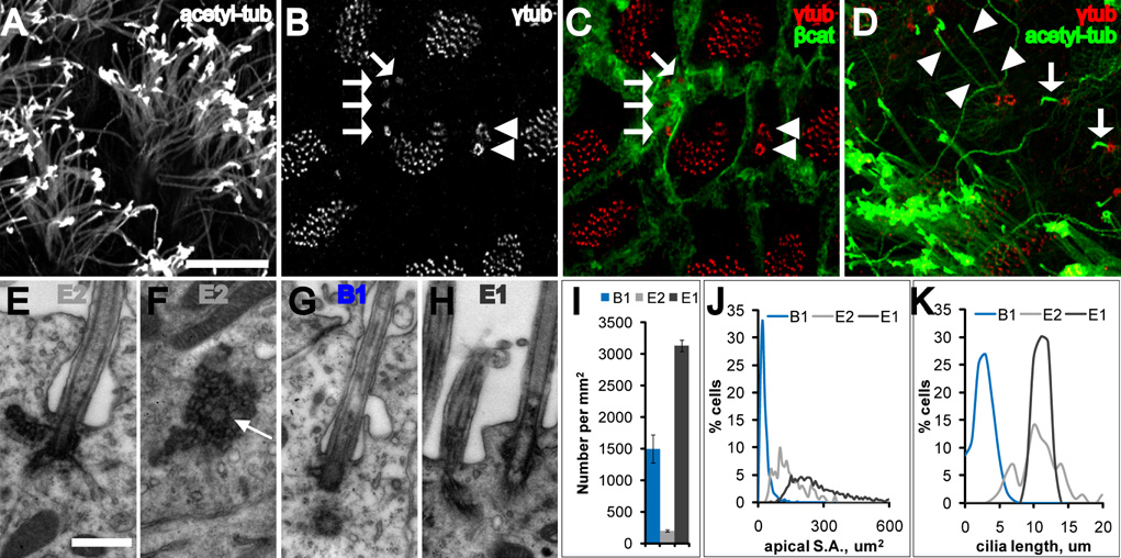
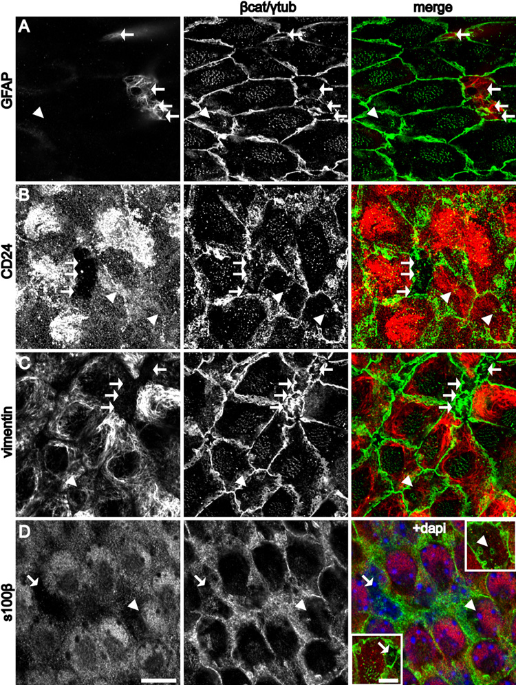
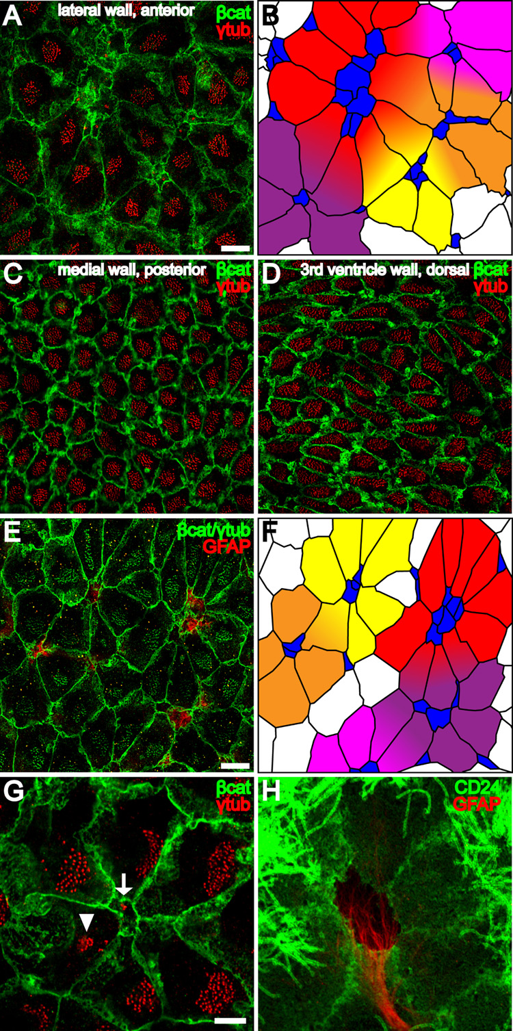
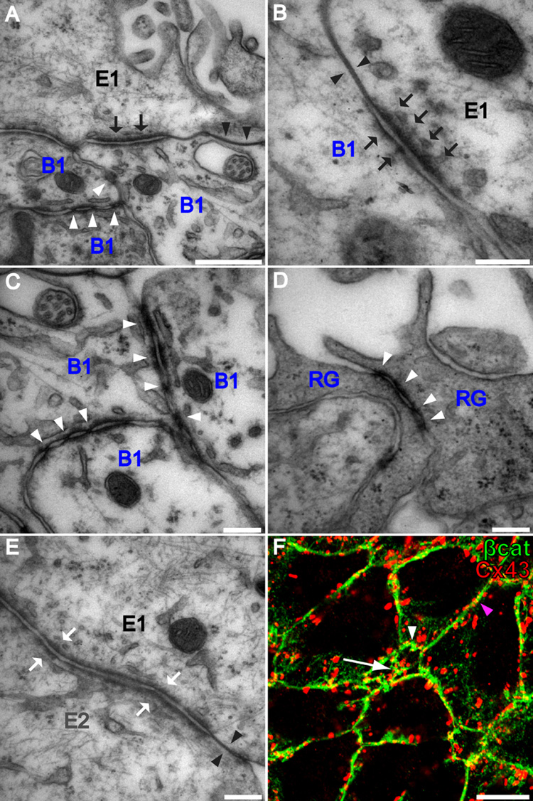
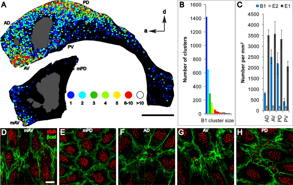
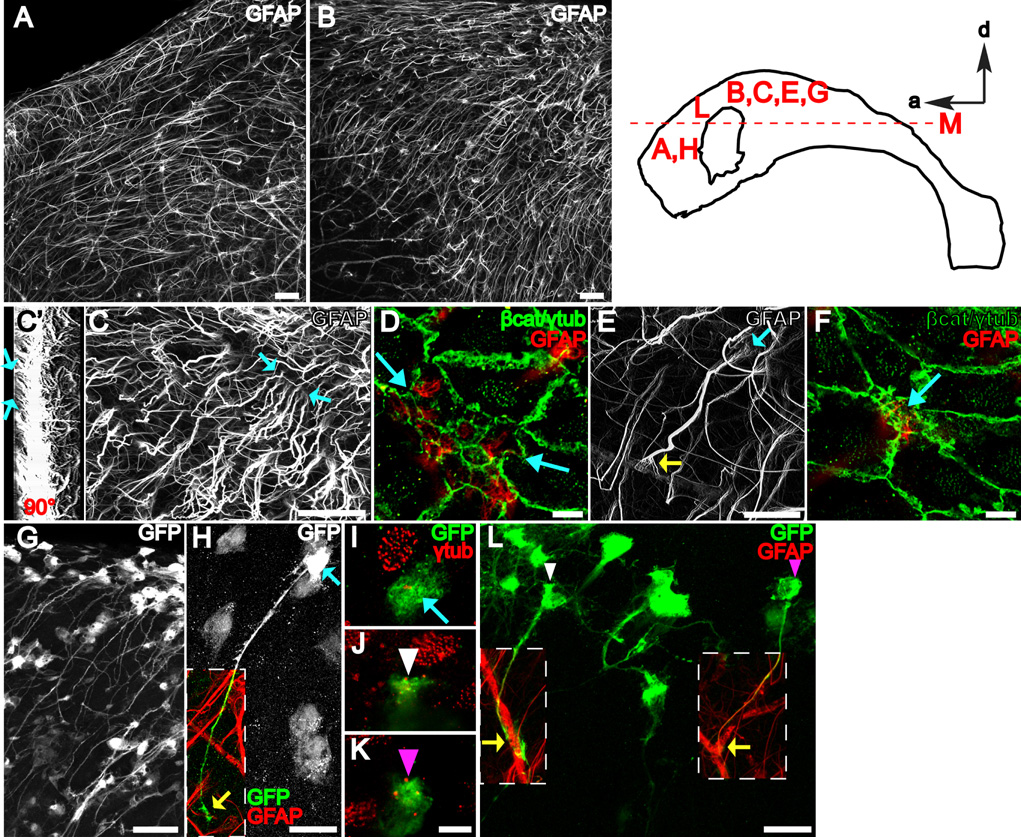
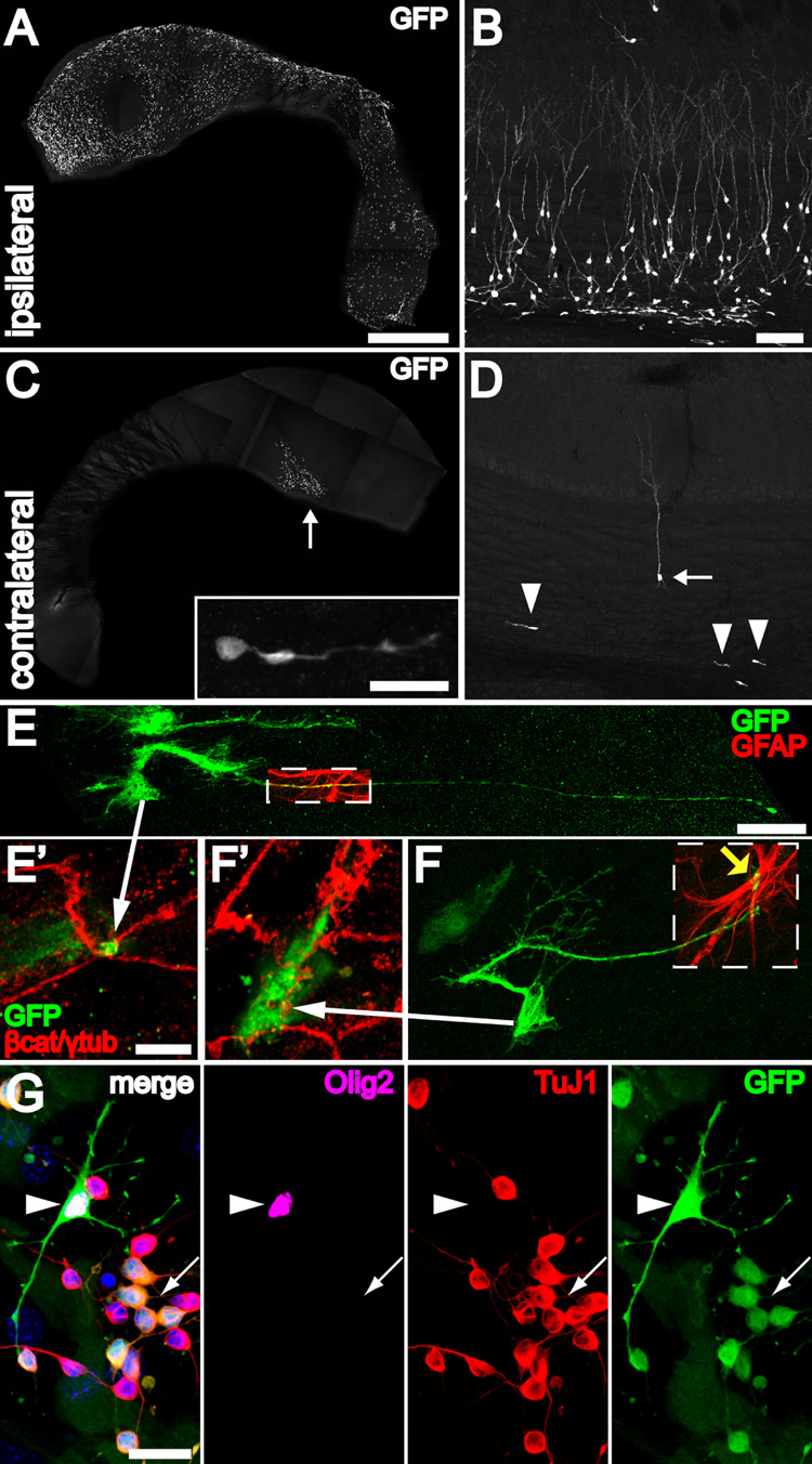
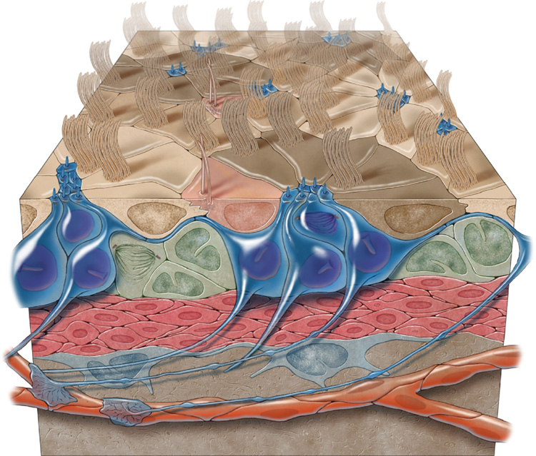
Neural stem cells (NSCs, B1 cells) are retained in the walls of the adult lateral ventricles but, unlike embryonic NSCs, are displaced from the ventricular zone (VZ) into the subventricular zone (SVZ) by ependymal cells. Apical and basal compartments, which in embryonic NSCs play essential roles in self-renewal and differentiation, are not evident in adult NSCs. Here we show that SVZ B1 cells in adult mice extend a minute apical ending to directly contact the ventricle and a long basal process ending on blood vessels. A closer look at the ventricular surface reveals a striking pinwheel organization specific to regions of adult neurogenesis. The pinwheel's core contains the apical endings of B1 cells and in its periphery two types of ependymal cells: multiciliated (E1) and a type (E2) characterized by only two cilia and extraordinarily complex basal bodies. These results reveal that adult NSCs retain fundamental epithelial properties, including apical and basal compartmentalization, significantly reshaping our understanding of this adult neurogenic niche.


Figures

Figure 1
Figure 1. Three cell types contact the LV
(A–D) Confocal images taken at the surface of wholemounts of the lateral wall of the LV. (A) Acetylated-tubulin staining reveals many long cilia of ependymal cells. Scale bar = 10 um (A–D). (B, C) γ-tubulin staining (B) together with β-catenin staining (C) reveals three cell types: E1 (many basal bodies), B1 (arrows indicate single basal body), and E2 (arrowheads indicate two basal bodies). (D) Three cell types are also distinguished by their cilia. B1 cells have a single short cilium (arrows) and E2 cells have 2 long cilia (arrowheads). (E, F) Electron micrographs from two serially reconstructed E2 cells. The central barrel of the basal body is cut transversely in (E) and in cross-section in (F) (arrow). Scale bar = 0.5 um (E–H). (G) Electron micrograph of the primary cilium, basal body and centriole of a B1 cell. Note the simpler B1 basal body compared with the large, lobular E2 basal body (F). Both B1 and E2 (E) cilia were invaginated in the apical membrane. (H) Electron micrograph of E1 cilia, which were not invaginated. (I) Density of B1, E1, and E2 cells on the lateral wall of the LV. Error bars show sem. (J) Histogram of apical surface area of B1, E1, and E2 cells. Data from 771 B1 cells, 1716 E1 cells, and 201 E2 cells from n=4 mice. (K) Histogram of cilia length of B1, E1, and E2 cells. Data from 361 B1 primary cilia, 55 ependymal cilia, and 106 E2 cilia from n=3 mice.
Figure 2
Figure 2. Complementary expression patterns of molecular markers at the apical surface of B1 and E1/E2 cells
(A–C, D insets) Confocal images at the surface of the wholemount and (D) was taken just below the surface. B1 cells (arrows) and E2 cells (arrowheads). (A) GFAP staining is present in B1, but not E1 or E2 cells. (B) CD24 is expressed in the cilia and on the apical surface of E1 and E2, but not B1 cells. (C) Vimentin is expressed at the apical surface of E1 and E2, but not B1 cells. (D) S100β expression is observed in the cell body of E1 and E2, but not B1 cells. Insets show the apical surface of the indicated B1 and E2 cells. Scale bar = 10 um (A–D). Inset: Scale bar = 5 um.
Figure 3
Figure 3. Pinwheel architecture of the ventricular surface in the adult neurogenic niche
(A) Confocal image of the surface of the lateral wall of the LV stained for γ-tubulin (red) and β-catenin (green) reveals pinwheels with centrally located B1 apical surfaces surrounded by ependymal cells. Scale bar = 10 um (A–D). (B) Tracing of the β-catenin+ membranes in (A) and color-coding of B1 cells (blue) and E1 cells (yellow, orange, red, purple and magenta) to illustrate 5 pinwheels. Some E1 cells occupy positions in more than one pinwheel. (C,D) Neither B1 apical surfaces nor pinwheels are observed in the posterior medial wall of the LV or in 3rd ventricle walls. (E) Confocal image of the surface of the lateral wall of the LV shows that pinwheels surround GFAP+ (red) B1 cells. β-catenin and γ-tubulin are in green. Scale bar = 10 um. (F) Color-coded tracing of the image in (E). (G) Confocal image demonstrating the diamond shape of E1 apical surfaces found in pinwheels. The polarized position of the basal body patch, found on the right side of each E1 surface in this image, is independent of the E1 surface geometry. A B1 apical surface is indicated in the center of the pinwheel (arrow) and an E2 apical surface (arrowhead) on the periphery. Scale bar = 5 um (G, H) (H) Pinwheel architecture revealed by GFAP and CD24, which label B1 and E1/E2 cells, respectively.
Figure 4
Figure 4. Apical intercellular junctional complexes differ at B1-B1, B1-E1, and E1–E2 junctions
(A) In addition to tight junctions (black arrowheads), B1 and E1 cells form asymmetric adherens junctions (black arrows). B1 cells form symmetric atypical adherens junctions with each other (white arrowheads). Scale bar = 500 nm. (B) Higher magnification of an asymmetric B1–E1 adherens junction (black arrows) and a B1–E1 tight junction (black arrowheads). Scale bar = 200 nm. (C) Higher magnification of atypical B1–B1 adherens junctions. Scale bar = 200 nm. (D) Radial glia (RG) in the neonatal VZ have apically localized symmetric atypical adherens junctions (white arrowheads) similar to those at B1–B1 junctions in the adult. Scale bar = 200 nm. (E) E1 and E2 cells form tight junctions (black arrowheads) and symmetric adherens junctions (white arrows) indistinguishable from those at E1-E1 junctions. Scale bar = 200 nm. (F) Confocal image of the surface of the lateral wall of the LV stained for Cx43 (red) and β-catenin (green) reveals that gap junctional components are present at B1-B1 (arrow), B1-E (white arrowhead), and E-E (magenta arrowhead) intercellular membranes. Scale bar = 10 um.
Figure 5
Figure 5. Surface maps of the walls of the LV reveal “hot spots” of B1 apical surfaces
(A) The position of B1 apical surfaces is indicated on the maps by color-coded circles depicting the number of B1 apical surfaces per cluster (1 to >10 cells/cluster). Hot spots were found in anterior-ventral (AV) and posterior-dorsal (PD) regions of the lateral wall and anterior-ventral (mAV) region of the medial wall. The adhesion point between lateral and medial walls is shown in gray. Scale bar = 1 mm. (B) Distribution of B1 cluster size. (C) Density of B1, E1, and E2 apical surfaces in 4 regions of the lateral wall in 3 animals. Regions quantified are indicated in (A) and Figure S13. Error bars show sem. (D–H) Representative confocal images from different regions of the surface of the lateral and medial walls stained for γ-tubulin and β-catenin. Scale bar = 5 um.
Figure 6
Figure 6. B1 cells with an apical ventricular contact have a long basal process terminating on blood vessels
The diagram of the lateral wall at the top right indicates where the images were taken. (A, B) Z-projections of confocal stacks (50 um thick) taken from wholemounts stained for GFAP. Because wholemounts are freshly dissected, secondary antibodies used against mouse anti-GFAP primary antibody also stain blood vessels that contain endogenous mouse IgGs. Scale bar = 50 um. (C) Z-projection of a higher power confocal stack (50 um thick) taken from a wholemount stained for GFAP. Blue arrows indicate the apical endings of long GFAP+ processes. Scale bar = 50 um. (C’) 3-dimensional reconstruction of the stack in (C) rotated 90°. Blue arrows indicate processes reaching the ventricular surface and correspond to blue arrows in C. (D) Confocal image of the surface (co-stained for γ-tubulin and β-catenin in green) in the region between the blue arrows in (C) reveals that the apical endings indicated in (C) belong to a group of 11 B1 cells with GFAP+ apical surfaces on the ventricle. Scale bar = 5 um. (E, F) The long basal process of a B1 cell with an apical ventricular contact (blue arrows) terminates on a blood vessel forming an endfoot (yellow arrow in E). Scale bar = 25 um (E), 5 um (F). (G) B1 cells with apical ventricular contact could be labeled by adeno-GFAPp:Cre virus injected into the LV of Z/EG mice. Scale bar = 50 um. (H) High power confocal z-stack projection reveals the complete morphology of a B1 cell: apical contact with the ventricle (blue arrows in H, I) and basal contact with blood vessels (yellow arrow in H). Scale bar = 25 um. (I–K) Confocal images of the ventricle-contacting apical surface of B1 cells corresponding to the color-coded arrow in (H) and arrowheads in (L). Scale bar = 5 um. (L) Adult B1 cells are derived from neonatal radial glia. Note the apical ventricular contact (white and magenta arrowheads in J, K, L) of two GFP+ B1 cells derived from neonatal radial glial labeling. Both cells have a long basal process contacting a blood vessel (yellow arrows). Scale bar = 25 um.
Figure 7
Figure 7. B1 cells with apical ventricular contact are neurogenic in vivo and differentiate into neurons, astrocytes and oligodendrocytes in vitro
(A) Injection of adeno-GFAPp:Cre into one LV of Z/EG mice results in many labeled ventricle-contacting B1 cells on the ipsilateral lateral wall. The wholemount images (A, C) were reconstructed from low power (1.3 mm × 1.3 mm) tiled confocal images. Scale bar = 1 mm (A, C) (B) 30 days after injection, many labeled neurons and neuroblasts can be found in the ipsilateral olfactory bulb. Scale bar = 100 um (B, D). (C) Virus that backfills the contralateral LV labels a few ventricle-contacting B1 cells on the contralateral side. Most of these labeled cells are close to the foramen of Monro (arrow). These labeled B1 cells give rise to neuroblasts (inset) in the contralateral SVZ, examined 30 days after injection. Inset: Scale bar = 20 um. (D) The few labeled ventricle-contacting B1 cells on the contralateral side gave rise 30 days later to neuroblasts (arrowheads) and neurons (arrow) in the contralateral OB. (E, F) B1 cells in the contralateral lateral wall have characteristic features of apical ventricular contact (arrows to E’, F’), GFAP expression (boxed regions), and a long basal process with endfeet on blood vessels (yellow arrow). Scale bar = 25 um. (E’, F’) Confocal images of the ventricle-contacting apical surface of B1 cells in (E, F). Scale bar = 5 um. (G) GFP-labeled B1 cells dissected from the contralateral wall could be passaged and differentiated in vitro into TuJ1+ neurons (arrow), Olig2+ oligodendrocytes (arrowhead), and adherent GFAP+ (not shown) astrocytes. Scale bar = 20 um.
Figure 8
Figure 8
3-dimensional model of the adult VZ neurogenic niche illustrating B1 cells (blue), C cells (green), and A cells (red). B1 cells have a long basal process that terminates on blood vessels (orange) and an apical ending at the ventricle surface. Note the pinwheel organization (light and dark brown) composed of ependymal cells encircling B1 apical surfaces. E2 cells are peach.

Comment in

  • The niche revealed.
    Currle DS, Gilbertson RJ. Currle DS, et al. Cell Stem Cell. 2008 Sep 11;3(3):234-6. doi: 10.1016/j.stem.2008.08.011. Cell Stem Cell. 2008. PMID: 18786409 No abstract available.

References

    1. Alvarez-Buylla A, Lim DA. For the long run: maintaining germinal niches in the adult brain. Neuron. 2004;41:683–686. - PubMed
    1. Alvarez-Buylla A, Nottebohm F. Migration of young neurons in adult avian brain. Nature. 1988;335:353–354. - PubMed
    1. Alvarez-Buylla A, Theelen M, Nottebohm F. Proliferation "hot spots" in adult avian ventricular zone reveal radial cell division. Neuron. 1990;5:101–109. - PubMed
    1. Bittman K, Owens DF, Kriegstein AR, LoTurco JJ. Cell coupling and uncoupling in the ventricular zone of developing neocortex. J Neurosci. 1997;17:7037–7044. - PMC - PubMed
    1. Breunig JJ, Silbereis J, Vaccarino FM, Sestan N, Rakic P. Notch regulates cell fate and dendrite morphology of newborn neurons in the postnatal dentate gyrus. Proc Natl Acad Sci USA. 2007;104:20558–20563. - PMC - PubMed

Publication types

LinkOut - more resources