. 2008 Sep 11;3(3):289-300.
doi: 10.1016/j.stem.2008.07.026.

Adult SVZ stem cells lie in a vascular niche: a quantitative analysis of niche cell-cell interactions

Affiliations

Adult SVZ stem cells lie in a vascular niche: a quantitative analysis of niche cell-cell interactions

Qin Shen et al. Cell Stem Cell. .

Abstract

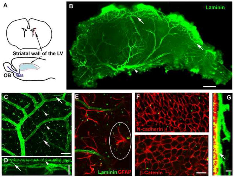
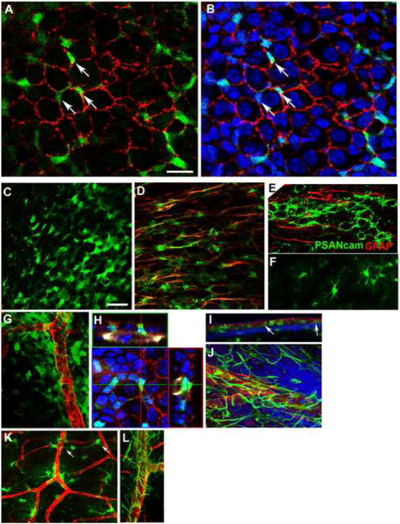
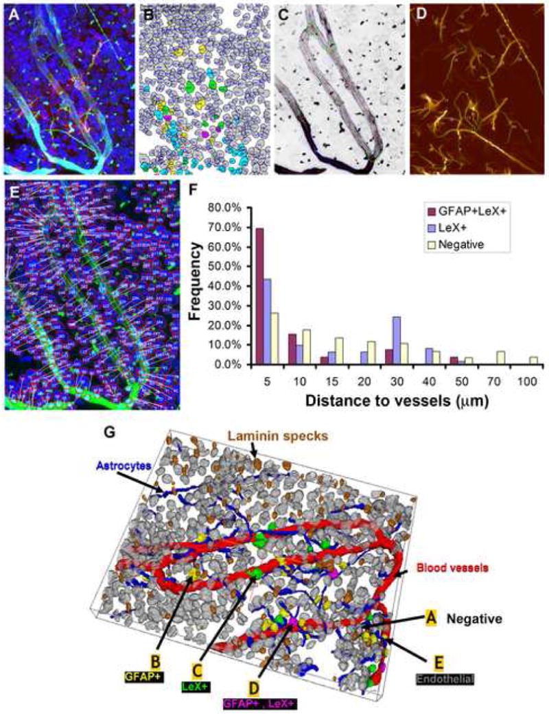
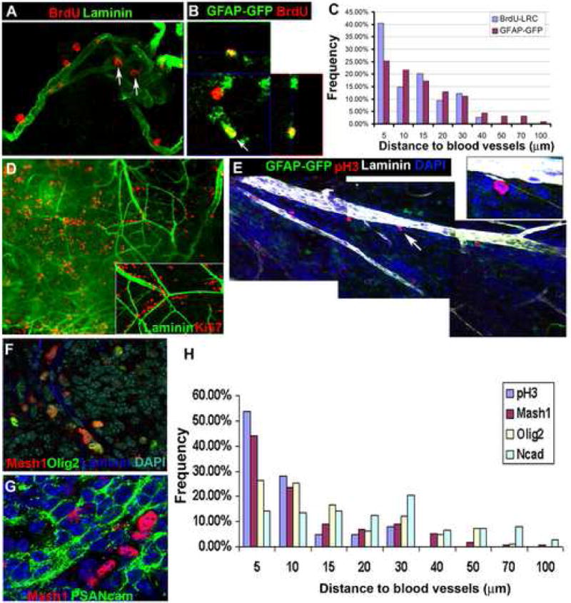
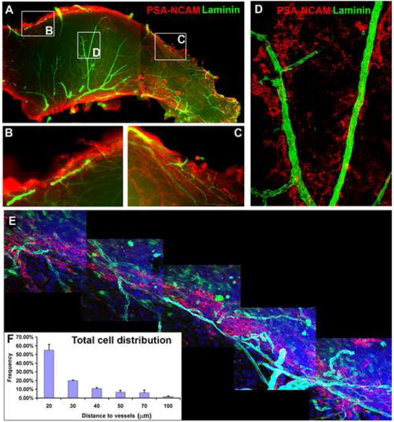
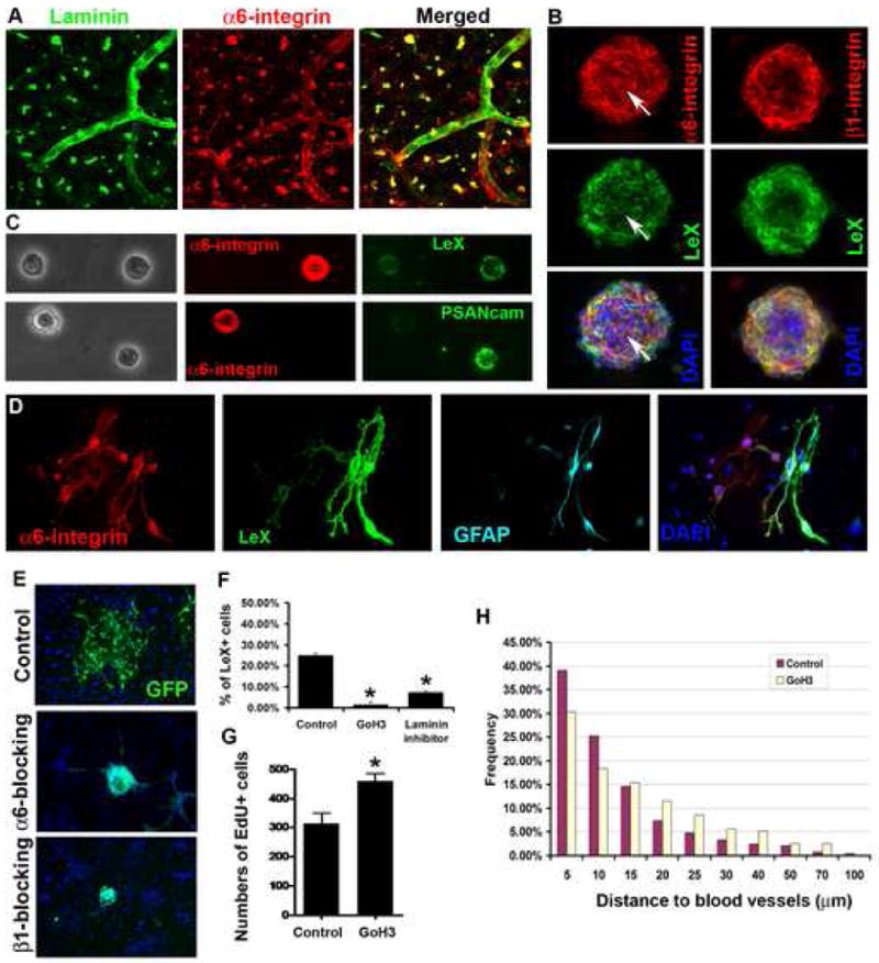
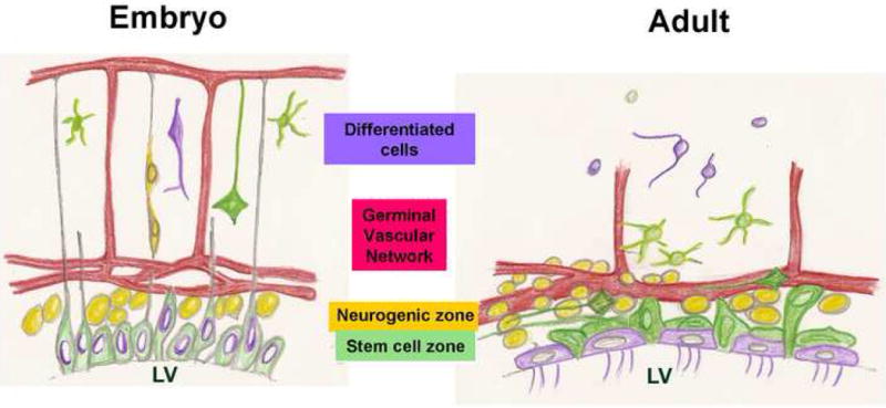
There is an emerging understanding of the importance of the vascular system within stem cell niches. Here, we examine whether neural stem cells (NSCs) in the adult subventricular zone (SVZ) lie close to blood vessels, using three-dimensional whole mounts, confocal microscopy, and automated computer-based image quantification. We found that the SVZ contains a rich plexus of blood vessels that snake along and within neuroblast chains. Cells expressing stem cell markers, including GFAP, and proliferation markers are closely apposed to the laminin-containing extracellular matrix (ECM) surrounding vascular endothelial cells. Apical GFAP+ cells are admixed within the ependymal layer and some span between the ventricle and blood vessels, occupying a specialized microenvironment. Adult SVZ progenitor cells express the laminin receptor alpha6beta1 integrin, and blocking this inhibits their adhesion to endothelial cells, altering their position and proliferation in vivo, indicating that it plays a functional role in binding SVZ stem cells within the vascular niche.


Figures

Figure 1
Figure 1. A dense plexus of blood vessels and associated Laminin+ structures in the SVZ
(A). SVZ wholemounts were dissected from the striatal wall of the lateral ventricle (red outline). OB, olfactory bulb; RMS, rostral migratory stream; LV, lateral ventricle. (B). Laminin staining shows the dense network of blood vessels in the SVZ wholemount (viewed from the ventricular surface; anterior, left and dorsal, top. Arrows: vessels running along the dorsal border of the SVZ. Arrowhead: a large vessel in the ventral aspect. (C-D). Laminin staining reveals blood vessels, Laminin specks (arrowheads) and fractones (arrows) (C, a projection image of confocal Z-stacks). A projection image generated with the z-axis as the turning axis (D) shows the superficial Laminin specks, the SVZ vessel network parallel to the ventricular surface, and the deeper vessel branches that delve down into the striatum. Top: LV surface, bottom: striatal side. (Arrowheads indicate Laminin specks near the surface; arrows indicate fractones). (E). Laminin specks were frequently contacted by GFAP+ processes, as circled. (F). N-cadherin (upper, red) or β-catenin (lower, red) shows the pavemented ependymal cell layer. Some Laminin specks (green) are seen in the ependymal layer. (G). Z-stacks of confocal images of an SVZ wholemount stained for N-cadherin (red) and Laminin (green) viewed from the z-axis. Left: ventricular surface, right: the striatal side. Note a section of vessel (arrow) immediately beneath the ependymal layer. Scale bars: B: 300 μm; C-D: 25 μm; F: 20 μm; G: 10 μm.
Figure 2
Figure 2. Three layers of distinct types of GFAP-GFP cells in the SVZ and their apposition to blood vessels
(A-B). Apical Type B cells are intercalated into the ependymal layer. (A) A single confocal scan of the SVZ wholemount from a GFAP-GFP mouse brain stained for N-cadherin (red) and GFP (green). (B) The same image shown in (A) with DAPI staining (blue) to show the inclusion of the nuclei of GFAP-GFP+ cells in the ependymal layer (3 examples indicated by arrows). (C-F). Imaging GFAP-GFP+ cells from the ventricular surface towards the striatum, single z-stack images at 5 μm (C), 10 μm (D) and 20 μm (F) from the ependymal layer. (C). Apical GFAP-GFP+ cells have a neuroepithelial-like morphology and form a layer mixed within and just beneath the ependymal layer. (D). Tangential GFAP-GFP+ cells with one or two long processes (stained for GFAP, red) are prevalent immediately beneath the apical layer; the aligned processes run anterior-posterior, in the direction of neuroblast migration (stained for PSA-NCAM, green in E). (F). Near the striatal border, GFAP-GFP cells are typical of more mature astrocytes with multiple processes. (G-L). GFAP-GFP+ cells and processes are closely apposed to blood vessels. (G). Numerous epithelial-like GFAP-GFP+ cells (green) wrap around blood vessels (Laminin, red). Many GFAP-GFP+ cell somas sit on the vessel walls. (H). Orthogonal sections of a confocal image showing an apical GFAP-GFP+ cell intercalated into the ependymal layer and contacting a blood vessel. The GFAP-GFP SVZ wholemount was stained for GFP (green), N-cadherin (red), Laminin (pseudocolored white), and DAPI (blue). (I). A projection image of Z-stacks (viewed from the z-axis) of a GFAP-GFP SVZ wholemount stained for GFP (green), N-cadherin (red), Laminin (blue) shows a layer of GFP+ cells between the ependymal layer and the SVZ blood vessels. Note that some GFP+ cells contact both the ventricle surface and the vessels (arrows). (J). Tangential SVZ astrocytes have long processes running along or between the blood vessels (GFAP, green, Laminin, red). (K). Deep GFAP-GFP+ cells are close to vessels with frequent endfeet touching blood vessels. Arrows indicate a cell with its soma on a vessel and its process touching another branch. (L). Deep astrocytic processes (GFAP, green) wrap around large vessels (Laminin, red). Scale bars: A-B: 20 μm; C, D, F: 50 μm.
Figure 3
Figure 3. GFAP+ and LeX+ cells near vessel walls in 3-D images of SVZ wholemounts
(A). 4-channel confocal image of SVZ wholemount stained for DAPI (blue), GFAP (green), Laminin (aqua), and LeX (red). (B). Results of automated delineation of cell nuclei in the DAPI channel, and automated cell classification indicated by color labels of nuclei (green, LeX+; yellow, GFAP+; purple, GFAP+ and LeX+; gray, all other nuclei (negative). (C). Automated vessel tracing computed on the Laminin channel (vessel center lines are displayed in red and surfaces in blue). (D). Automated tracing of astrocyte processes (green). (E). Illustration of the method for computing distances of nuclear centroids to the nearest vessel segment. (F). Histogram of pooled validated data from 6 images showing the distances of cells that were double LeX+GFAP+ or Lex+GFAP- to vasculature as compared to negative cells. (G). 3D rendering of the segmentation and classification results. The cell nuclei are colored as in (B), vessel surfaces in red, astrocyte processes in blue, and Laminin specks brown. (labels A – E indicate cells that are negative, GFAP+, LeX+, GFAP+LeX+, and endothelial).
Figure 4
Figure 4. Neurogenesis occurs around the vessel network in the adult SVZ
(A-C). Adult mice were given 5 daily BrdU injections then a 24 day chase to reveal LRCs. (A). A confocal image of the SVZ showing BrdU LRC (red) proximity to blood vessel (Laminin+, green); some BrdU+ cells are in a niche formed by a knot of vessels (arrows). (B). An orthogonal section of confocal z-stacks. Arrow: a BrdU-retaining cell (red) that is positive for GFAP-GFP. 25.0% of LRCs were found to be GFAP-GFP+ after GFP staining, similar to the 38% observed using a 6 week chase period (Tazavoie et al, 2008). The identity of GFAP-ve LRCs remains to be determined. (C). Histogram of distance of LRCs and GFAP-GFP cells to vessels (Data from 13 images derived from 4 SVZ wholemounts). (D). A low power image of an SVZ wholemount stained for the cell proliferation marker Ki67 (red) and Laminin (green). Ki67+ cells are frequently aligned next to blood vessels (arrow indicates the area shown inset in a higher magnification confocal image). (E). A montage of single Z-sections of confocal images of a GFAP-GFP SVZ wholemount stained for pH3+ cells (red), Laminin (pseudocolored white), and DAPI (blue). PH3+ cells are adjacent to the vessels. Note one positive cell (arrow and inset) next to a Laminin+ fractone. (F). Double-labeling of Mash1 (red) and Olig2 (green). (G). Mash1+ cells (red) are near their lineal progeny PSA-NCAM+ Type A cells. (H). A histogram of the distance to vessels of cells that are pH3+, Mash1+, Olig2+ and N-cadherin+. Most pH3+ cells are within 10 μm of the nearest vessel, Mash1+ cells are similarly close while Olig2+ cells are more widely distributed around the vessels. The distances of N-cadherin+ ependymal cells were plotted as a reference, and range from 5 μm-100 μm. Data pooled from 5 randomly chosen images.
Figure 5
Figure 5. Neuroblasts migrate in a chain along blood vessels
(A-D). Low power image of SVZ wholemount stained for PSA-NCAM (red) and Laminin (green); Left, anterior and top, dorsal. High magnification images of boxed areas are shown in (B-D). Chains of migrating PSA-NCAM+ neuroblasts are aligned along blood vessels in the anterior dorsal region (B), the posterior region (C) and some occur in the central SVZ (D). (E). A montage of confocal images showing approximately 1 mm of the dorsal region of an SVZ wholemount derived from a GFAP-GFP brain stained for PSA-NCAM (red), Laminin (cyan) and DAPI (blue). A vessel complex travels along and within the neuroblast chain. (F). Histogram showing the relationship between SVZ cell distribution and blood vessels. More than 50% of cells are within 20 μm of blood vessels. Data represents mean ± SEM from 3 SVZ wholemounts (3-5 images per wholemount).
Figure 6
Figure 6. α6β1 integrin is expressed in adult SVZ cells and required for SVZ cells to adhere to endothelial cells
(A-D). α6 and β1 integrin expression in the SVZ and NSCs. (A). An SVZ wholemount stained for α6 integrin (red) and Laminin (green). α6 integrin staining is seen on laminin specks and blood vessels, as well as on SVZ cells. (B). α6 and β1 integrins detected in SVZ neurospheres and on LeX+ progenitor cells (green), DAPI in merged images. Arrows point to a dividing cell double-positive for α6 integrin and LeX. (C). Acutely fixed single SVZ cells show strong α6 integrin expression in a subpopulation of LeX positive cells, but weak or no staining in PSA-NCAM+ neuroblasts. (D). A 3-day SVZ clonal culture, the α6 integrin+ cells are LeX+GFAP+. (E). After treatment with blocking antibodies to α6 or β1 integrin, SVZ neurospheres derived from adult GFP mice spread poorly on endothelial cells. (F). Blocking with anti-α6 integrin antibody (GoH3) or laminin binding inhibitor peptide inhibits adhesion to endothelial monolayers. Data, Mean±SEM (n=2 experiments. ANOVA, Bonferroni test *P<0.005). (G). Significantly increased proliferation (EdU-incorporated cells) in the dorsal SVZ after 6-day in vivo infusion of anti-α6 integrin. Data, Mean±SEM (n=3 SVZ wholemounts. *P<0.05, t-test). (H). S-phase cells are significantly further from blood vessel surfaces after 6-day in vivo infusion of GoH3 antibody (Mann-Whitney test, P<0.001, n=234 cells for GoH3, n=246 cells for control group).
Figure 7
Figure 7. Summary model comparing the vascularization of adult and embryonic germinal zones
In the embryo, an apical layer of stem cells produces an actively proliferating SVZ which generates neurons that migrate towards the pia guided by radial glia. Coincident with neurogenesis, the vasculature grows from the pial surface towards the germinal cells, a source of VEGF (Breier et al., 1992) and form a plexus at the germinal zones, parallel to the ventricular surface (Shen et al., 2004; Strong, 1964). The adult SVZ has an essentially similar structure: Apical Type B cells, believed to include the SVZ stem cells, are intercalated into the ependymal layer and directly contact the ventricle. Just subjacent is an SVZ of active proliferation, differentiation and migration, including Mash1+ and Olig2+ Type C progenitors, PSA-NCAM+ Type A neuroblasts and Tangential Type B cells. The adult germinal zone is intimately associated with an SVZ vascular plexus.

Comment in

  • The niche revealed.
    Currle DS, Gilbertson RJ. Currle DS, et al. Cell Stem Cell. 2008 Sep 11;3(3):234-6. doi: 10.1016/j.stem.2008.08.011. Cell Stem Cell. 2008. PMID: 18786409 No abstract available.

References

    1. Alvarez-Buylla A, Garcia-Verdugo JM, Mateo AS, Merchant-Larios H. Primary neural precursors and intermitotic nuclear migration in the ventricular zone of adult canaries. J Neurosci. 1998;18:1020–1037. - PMC - PubMed
    1. Alvarez-Buylla A, Lim DA. For the long run: maintaining germinal niches in the adult brain. Neuron. 2004;41:683–686. - PubMed
    1. Baker KL, Daniels SB, Lennington JB, Lardaro T, Czap A, Notti RQ, Cooper O, Isacson O, Frasca S, Jr, Conover JC. Neuroblast protuberances in the subventricular zone of the regenerative MRL/MpJ mouse. J Comp Neurol. 2006;498:747–761. - PubMed
    1. Breier G, Albrecht U, Sterrer S, Risau W. Expression of vascular endothelial growth factor during embryonic angiogenesis and endothelial cell differentiation. Development. 1992;114:521–532. - PubMed
    1. Brightman MW. The distribution within the brain of ferritin injected into cerebrospinal fluid compartments. I. Ependymal distribution. J Cell Biol. 1965;26:99–123. - PMC - PubMed

Publication types

MeSH terms

Substances