. 2008 Nov 1;323(1):41-52.
doi: 10.1016/j.ydbio.2008.08.011. Epub 2008 Aug 22.

The chromatin remodelers ISWI and ACF1 directly repress Wingless transcriptional targets

Affiliations

The chromatin remodelers ISWI and ACF1 directly repress Wingless transcriptional targets

Yan I Liu et al. Dev Biol. .

Abstract

The highly conserved Wingless/Wnt signaling pathway controls many developmental processes by regulating the expression of target genes, most often through members of the TCF family of DNA-binding proteins. In the absence of signaling, many of these targets are silenced, by mechanisms involving TCFs that are not fully understood. Here we report that the chromatin remodeling proteins ISWI and ACF1 are required for basal repression of WG target genes in Drosophila. This regulation is not due to global repression by ISWI and ACF1 and is distinct from their previously reported role in chromatin assembly. While ISWI is localized to the same regions of Wingless target gene chromatin as TCF, we find that ACF1 binds much more broadly to target loci. This broad distribution of ACF1 is dependent on ISWI. ISWI and ACF1 are required for TCF binding to chromatin, while a TCF-independent role of ISWI-ACF1 in repression of Wingless targets is also observed. Finally, we show that Wingless signaling reduces ACF1 binding to WG targets, and ISWI and ACF1 regulate repression by antagonizing histone H4 acetylation. Our results argue that WG signaling activates target gene expression partly by overcoming the chromatin barrier maintained by ISWI and ACF1.


Figures

Fig. 1
Fig. 1
ISWI and ACF1 repress Wg targets in cultured cells. (A) Kc cells were treated with control media or Wg-CM for 5 hours prior to harvest. Transcript levels of nkd, Notum and hth were measured by qRT-PCR and results were normalized to β-tubulin56D expression. Wg-CM significantly induced the expression of all three genes. (B) Western blot analysis of ISWI and ACF1 in control or corresponding RNAi-treated cells. iswi RNAi and acf1 RNAi dramatically reduced ISWI and ACF1 expression, respectively. Arrows indicate the positions of ISWI and ACF1 proteins, and asterisks indicate nonspecific bands. (C) Derepression of nkd, Notum and hth by iswi or/and acf1 RNAi. Kc cells were treated with the indicated dsRNAs as described in Materials and methods, and transcripts of Wg targets were measured by qRT-PCR. Results were normalized to the average of β-tubulin56D, arm and TCF expression. (D) Two genes adjacent to nkd locus, Mkp3 and CG18135, as well as p53 were not derepressed by iswi, acf1 RNAi. The same normalization strategy was used as in (C). (E) arm RNAi did not affect the derepression of nkd by iswi, acf1 RNAi. (F) Activation of nkd expression by Wg-CM is enhanced by knockdown of iswi and acf1. Each bar in this figure represents the mean (+ S.E.) of duplicate cultures with duplicate qRT-PCR reactions. All experiments have been performed at least three separate times with similar results.
Fig. 2
Fig. 2
Loss of iswi results in an expansion and/or derepression of Wg targets in wing imaginal discs. Confocal images of wing imaginal discs of late third instar larva stained for Wg (C, G, I, K), Notum-lacZ (B, F), and nkd-lacZ (J, L). (A–H) Mitotic clones of iswi1 were marked by the absence of GFP (green in A and E). Notum-lacZ expression is expanded in clones along the Wg expression domain (A–D, 95% penetrance, n=39), and is derepressed in clones far away from the D/V boundary (E–H, 64% penetrance, n=22). (I-L) nkd-lacZ expression is expanded in wing discs from iswi1/iswi2 transheterozygotes (L, 92% penetrance, n=24) while Wg expression is unaffected (K).
Fig. 3
Fig. 3
iswi mutant cells do not affect several non-Wg targets in wing imaginal discs. Confocal images of wing imaginal discs of late third instar larva stained for Dpp-lacZ (B) and Spalt (D, E). All iswi clones examined show normal Dpp-lacZ expression (B, n=24) and the width of the Spalt expression domain was unaffected in iswi1/iswi2 transheterozygotes (E, n=12).
Fig. 4
Fig. 4
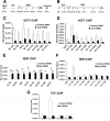
ACF1 and ISWI have different distributions on Wg target genes. (A, B) Schematic diagrams of the nkd and hth loci with the illustrated sites used for ChIP analysis. Arrows indicate the TSSs. The numbers in parentheses indicate the distance (in kb) from the TSS. N(−10) and N(+5) indicate the location of the two WREs of nkd. H(+16) is an area of hth bound by TCF. (C, D) ChIP analysis shows ACF1 binding to nkd and hth. Kc cells were treated with control dsRNA or acf1 dsRNA for 6 days before they were harvested for ChIP analysis. For nkd, ACF1 binds to a broad region as well as the genes adjacent to it (C). For hth, ACF1 also bound broadly but there was a three-fold enrichment of ACF1 binding at H(+16) that was reproducibly observed (D). (E, F) ChIP analysis of ISWI binding to nkd and hth. Assays were performed as for ACF1. There is no detectable ISWI binding (i.e., signal significantly higher than that in iswi depleted cells) on nkd (E), but there is ISWI enrichment at H(+16) on hth (F). (G) ChIP with TCF antisera shows specific binding of TCF to H(+16). Each bar in the figure represents the mean (± S.E.) of duplicate cultures with duplicate qPCR reactions. All experiments were performed three separate times with similar results except for the TCF ChIP, which was performed twice.
Fig. 5
Fig. 5
Relationship between binding of ACF1, ISWI and TCF to Wg target gene chromatin. (A, B) ACF1 binding is iswi-dependent. Kc cells were depleted of iswi for six days before processing for ACF1 ChIP. Marked reduction in binding was observed across the nkd and hth loci. (C) iswi knockdown does not reduce cellular ACF1 levels. Cells were treated with the indicated dsRNA for six days and processed for Western blot analysis with ACF1 antisera. Depletion of iswi does not affect ACF1 protein levels. (D) ISWI binds to hth independently of acf1. Depletion of acf1 does not cause a significant and reproducible reduction of ISWI binding to N(+5) or H(+16). (E, F) Depletion of TCF has no effect on ACF1 (E) or ISWI (F) binding to nkd and hth. The efficiency of acf1 and TCF RNAi in panels D–F was confirmed by Western blot (data not shown). (G) Depletion of iswi and acf1 reduces TCF binding to nkd and hth. Depletion of acf1 alone also reduces TCF binding to nkd and hth, but to a lesser degree compared to iswi, acf1 double knockdown cells (data not shown). (H) iswi, acf1 RNAi has no obvious effect on TCF expression. Western blot of TCF protein was performed on extracts treated with the indicated dsRNAs. TCF RNAi significantly reduced TCF protein levels but iswi, acf1 RNAi did not. Asterisks indicate nonspecific bands. (I) Depletion of TCF and iswi cooperately derepresses nkd and hth transcript levels in Kc cells. All experiments have been performed three separate times (except for the ISWI ChIP, which was performed twice) with similar results. The data in panels E and G are expressed as relative input because the absolute % input values differed from the nkd and hth loci.
Fig. 6
Fig. 6
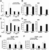
ISWI and ACF1 repress Wg targets independent of post-mitotic chromatin assembly. (A) Hydroxyurea (HU) effectively blocks cell division. Kc cells incubated with control or iswi and acf1 dsRNA for four days were treated with H2O or 5mM HU for an additional 48 hours. Cells stopped dividing, judged by cell number, upon HU treatment. In the control group, iswi, acf1 RNAi decreases the cell division rate. A similar decrease in cell division was also observed with acf1 RNAi, but not with iswi RNAi (data not shown). (B) Derepression of nkd and hth by iswi, acf1 RNAi is not abolished after HU treatment. Same experimental conditions were used as in (A), and transcript levels of nkd and hth were measured by qRT-PCR. (C) Aphidicolin (Aph) also inhibits cell division. Kc cells incubated with control dsRNA or iswi/acf1 dsRNA for four days were treated with DMSO or 25μM Aph for an additional 24 hours. Effective blockage of cell division was seen in Aph treated cells. (D) iswi, acf1 RNAi still derepresses nkd and hth after Aph treatment. (E, F) iswi, acf1 RNAi does not significantly affect H4 binding to the nkd and hth genes. ChIP analysis for pan-H4 was performed in control dsRNA or iswi/acf1 dsRNA treated cells. Multiple sites including the nkd WREs and TCF binding region of hth were tested for H4 binding, and no obvious change was observed between control RNAi and iswi/acf1 RNAi. Data shown were the means of duplicates (± S.E.), and all experiments have been performed two separate times with similar results.
Fig. 7
Fig. 7
ACF1 binding to Wg targets is modestly reduced by Wg signaling. (A, B) Cells were treated with control media or Wg-CM for 5 hours before harvesting for ACF1 ChIP analysis. A modest decrease of ACF1 binding was observed across the nkd and hth loci. (C) Less ACF1 binds to AcH4 on hth upon Wg-CM treatment. Cells treated with Wg-CM for 5 hours underwent ChIP with either α-H4 antibody or α-AcH4 antibody, followed by a secondary ChIP with α-ACF1 antibody. The α-AcH4 antibody recognizes acetylated K5/8/12/16 on histone H4. The re-ChIP signal was normalized to the eluted solution from the first immunoprecipitate, termed the 2nd input. (D) ISWI and ACF1 antagonize AcH4 levels on hth in the absence of Wg signaling. Cells were treated with control dsRNA or iswi, acf1 dsRNA for six days before harvested for AcH4 ChIP analysis. An increase of AcH4 on hth, most prominently at the site bound by TCF, was observed. In general, data represent the means of duplicates (± S.E.), and all experiments have been performed at least two separate times with similar results.

Similar articles

Cited by

References

    1. Akiyama H, Lyons JP, Mori-Akiyama Y, Yang X, Zhang R, Zhang Z, Deng JM, Taketo MM, Nakamura T, Behringer RR, McCrea PD, de Crombrugghe B. Interactions between Sox9 and beta-catenin control chondrocyte differentiation. Genes Dev. 2004;18:1072–1087. - PMC - PubMed
    1. Alenghat T, Yu J, Lazar MA. The N-CoR complex enables chromatin remodeler SNF2H to enhance repression by thyroid hormone receptor. Embo J. 2006;25:3966–3974. - PMC - PubMed
    1. Azpiazu N, Morata G. Function and regulation of homothorax in the wing imaginal disc of Drosophila. Development. 2000;127:2685–2693. - PubMed
    1. Bachman N, Gelbart ME, Tsukiyama T, Boeke JD. TFIIIB subunit Bdp1p is required for periodic integration of the Ty1 retrotransposon and targeting of Isw2p to S. cerevisiae tDNAs. Genes Dev. 2005;19:955–964. - PMC - PubMed
    1. Barolo S, Carver LA, Posakony JW. GFP and beta-galactosidase transformation vectors for promoter/enhancer analysis in Drosophila. Biotechniques. 2000;29:726–732. - PubMed

Publication types

MeSH terms

LinkOut - more resources