Comparative Study
. 2008 Oct 15;156(3):498-514.
doi: 10.1016/j.neuroscience.2008.08.013. Epub 2008 Aug 19.

Robust axonal growth and a blunted macrophage response are associated with impaired functional recovery after spinal cord injury in the MRL/MpJ mouse

Affiliations
Comparative Study

Robust axonal growth and a blunted macrophage response are associated with impaired functional recovery after spinal cord injury in the MRL/MpJ mouse

S K Kostyk et al. Neuroscience. .

Abstract

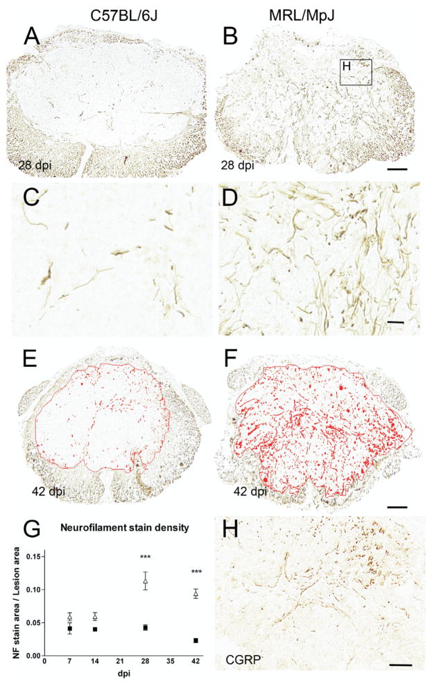
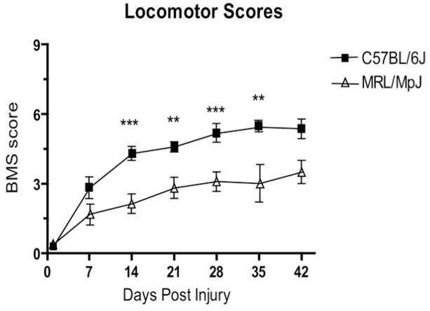
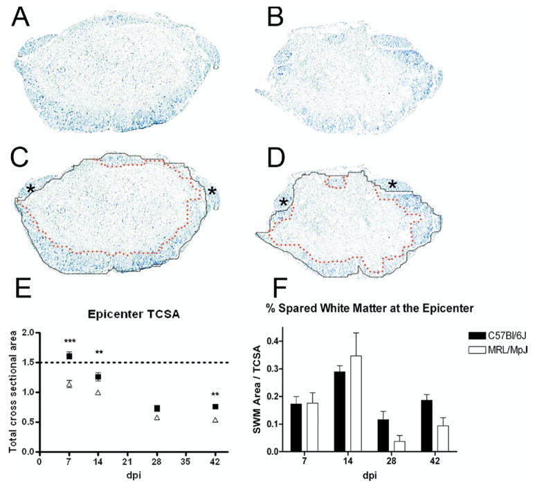
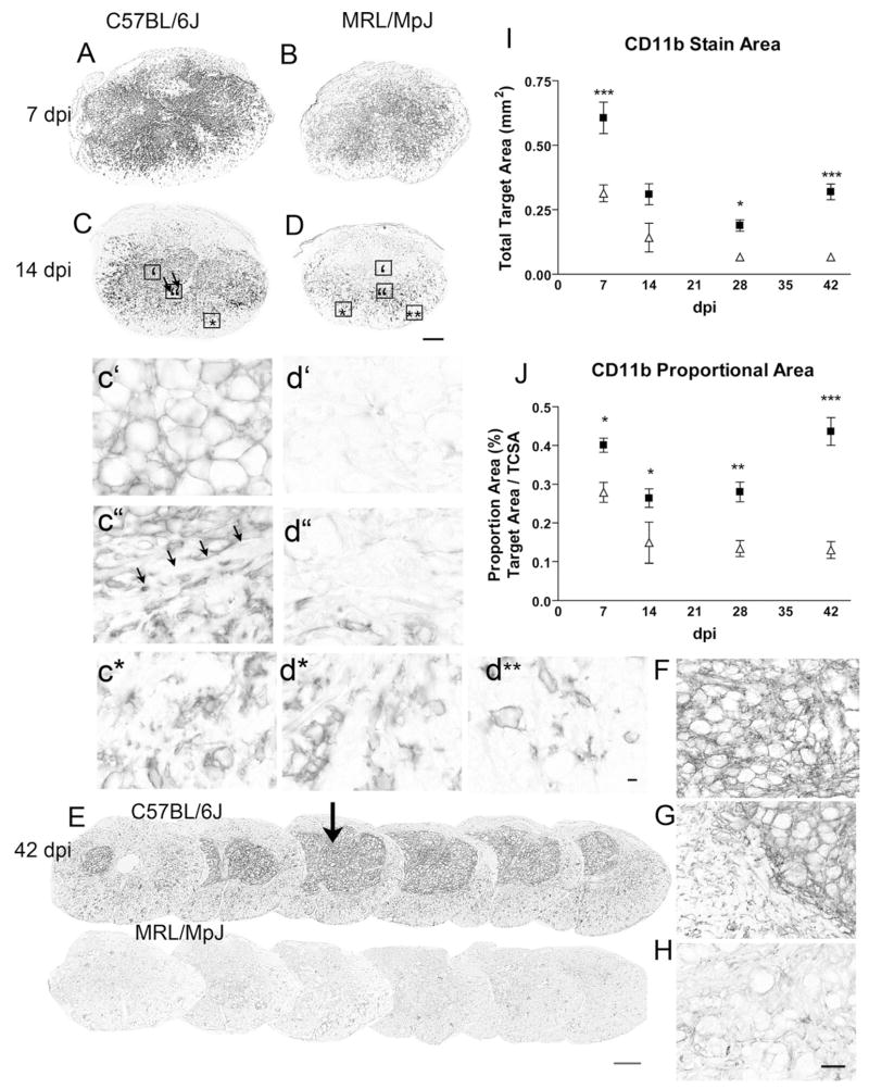
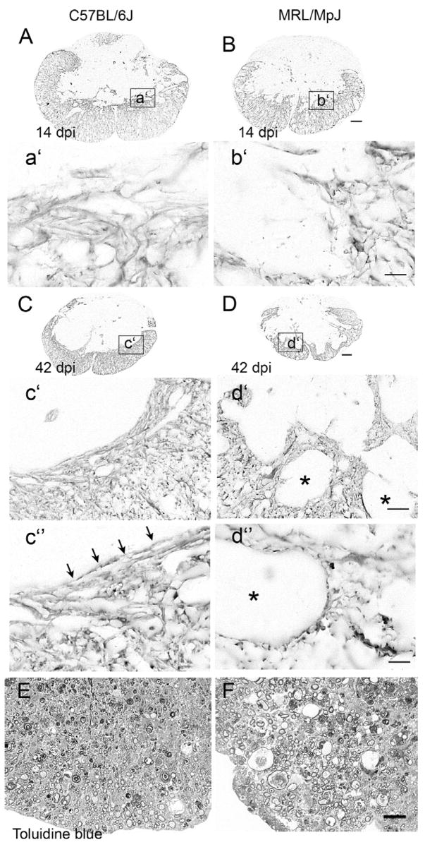
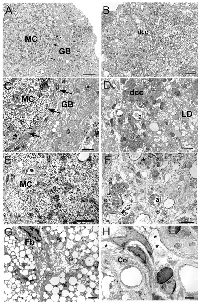
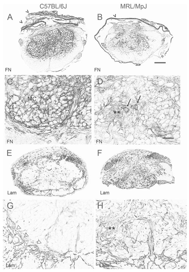
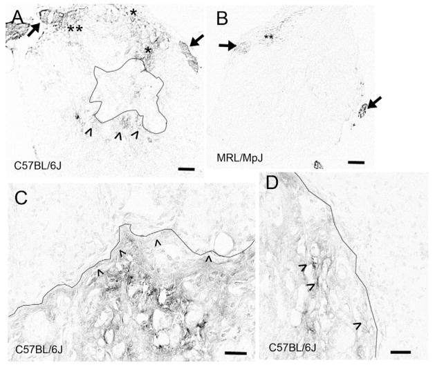
Spinal cord injury (SCI) in mammals leads to a robust inflammatory response followed by the formation of a glial and connective tissue scar that comprises a barrier to axonal regeneration. The inbred MRL/MpJ mouse strain exhibits reduced inflammation after peripheral injury and shows true regeneration without tissue scar formation following an ear punch wound. We hypothesized that following SCI, the unique genetic wound healing traits of this strain would result in reduced glial and connective tissue scar formation, increased axonal growth, and improved functional recovery. Adult MRL/MpJ and C57BL/6J mice were subjected to a mid-thoracic spinal contusion and the distribution of axon profiles and selected cellular and extracellular matrix components was compared at 1, 2, 4 and 6 weeks post-injury. Recovery of hind-limb locomotor function was assessed over the same time period. The MRL/MpJ mice exhibited robust axon growth within the lesion, beginning at 4 weeks post-injury. This growth was accompanied by reduced macrophage staining at 1, 2, 4 and 6 weeks post-injury, decreased chondroitin sulfate proteoglycan staining at 1-2 weeks and increased laminin staining throughout the lesion at 2-6 weeks post-injury. Paradoxically, the extent of locomotor recovery was impaired in the MRL/MpJ mice. Close examination of the chronic lesion site revealed evidence of ongoing degeneration both within and surrounding the lesion site. Thus, the regenerative genetic wound healing traits of the MRL/MpJ mice contribute to the evolution of a lesion environment that supports enhanced axon growth after SCI. However, this response occurs at the expense of meaningful functional recovery.


Figures

Fig. 1
Fig. 1
NF immunostaining reveals extensive axonal growth within the center of a contusion lesion in MRL/MpJ, but not C57BL/6J mice. Transverse sections through the lesion epicenter from C57BL/6J (A, C, E) and MRL/MpJ (B, D, F) specimens taken at 28 (A–D) and 42 (E, F) dpi, no differences were seen at 7 or 14 dpi. C and D are enlargements from the center of the lesion in specimens from A and B, respectively. (E, F) Axon profiles were quantified by identifying the density threshold for positive staining in the lesion site as indicated by the red binary overlay. Scale bar=100 μm (B, F for A, B and E, F); 20 μm (D for C, E). (G) The PA occupied by axons is greater in MRL/MpJ mice (open triangles) than in C57BL/6J mice (black squares). Two-way ANOVA indicated significant effects of strain (P<0.001; F=77.6, df=1), dpi (P<0.001; F=6.9, df=3) and an interaction effect (P<0.001; F=8.96, df=3) with MRL/MpJ greater at 28 and 42 dpi (P<0.001). (H) CGRP immunoreactivity from a section close to the NF stained section in B. A subset of the axons in the 28-day MRL/MpJ lesion exhibits CGRP-immunoreactivity (scale bar=10 μm).
Fig. 2
Fig. 2
Behavioral recovery after SCI is worse in MRL/MpJ mice (open triangles) than C57BL/6J mice (black squares) (two-way ANOVA strain P<0.001; dpi P<0.001; interaction P<0.05; post hoc Bonferroni P<0.001 at 14, 28 dpi, P<0.01 at 21, 35 dpi).
Fig. 3
Fig. 3
LFB-stained sections from the lesion site in C57BL/6J (A, C) and MRL/MpJ (B, D) mice at 42 dpi reveal subtle differences in the evolution of the lesion epicenter using classical white matter sparing measures. (A, B) Sections from both strains show a similar injury pattern including a central region of damage and a rim of peripheral SWM (F). (C, D) The same specimens with outlines to illustrate details of analysis. The TCSA of the section is outlined in black; asterisks identify dorsal roots, which were excluded from analysis. The inner rim of peripheral SWM is outlined in red. Scale bar=200 μm (A–D). (E) TCSA at the epicenter was smaller in MRL/MpJ mice (white triangles) than in C57BL/6J mice (black squares) at 7, 14 and 42 dpi. Dotted line depicts TCSA of uninjured spinal cord. Two-way ANOVA, P<0.0001 for strain and dpi; interaction P<0.01; Bonferroni post hoc ** P<0.01, *** P<0.001. (F) SWM at the rim expressed as a percent of cross-sectional area over time. Two-way ANOVA revealed an effect of dpi (P<0.001), but no effect of strain (P>0.05) or interaction (P>0.05). For interpretation of the references to color in this figure legend, the reader is referred to the Web version of this article.
Fig. 4
Fig. 4
CD11b (Mac-1) immunoreactivity was diminished in MRL/MpJ mice at all time points examined. Corresponding sections from the epicenter of C57BL/6J (A, C, E–G) and MRL/MpJ (B, D, E, H) specimens were stained simultaneously. (A) Macrophages are found throughout the lesion center in both strains at 7 dpi. (B) The area and intensity of Mac-1 staining were lower in MRL/MpJ mice; (C) 14 dpi in C57BL/6J mice. Boxes indicate regions enlarged below. The lesion contains a large area of Mac-1 staining, with rounded MCs throughout the center (c′), and a thin rim devoid of Mac-1 staining bordering the clusters (c″ arrows). Below this rim, activated Mac-1 positive cells exhibit heterogeneous morphologies suggesting a microglial origin. (D) Little to no staining was found in the lesion center in the MRL/MpJ mice at 14 dpi (d″), while elongated Mac-1+ cells line the borders (d″). Both strains have similar microglial distribution and activation in the rim of white matter (c*, d*, d**); (E) 42 dpi. Mac-1-stained sections spaced 300 μm apart through the lesion site in representative animals at 42 dpi illustrating the marked reduction in macrophage staining in MRL/MpJ mice at this chronic time point. The lesion epicenter section is marked by the arrow. (F–H) Enlargements from the sections stained at 42 dpi. (F) Center of a C57BL/6J lesion. (G) Border of the lesion center in C57BL/6J specimen. (H) Center of an MRL/MpJ lesion. (E, I, J) Time course of total (I) and proportional (J) area measures of Mac-1 (CD11b) labeled-macrophage/microglial profiles at the epicenter section in C57BL/6J and MRL/MpJ specimens Two-way ANOVA effect of dpi and strain for both measures (P<0.0001), differences between strains at each time point marked with asterisks (* P<0.05, ** P<0.01, *** P<0.0001). Scale bar=200 μm A–E; d**: Scale bar=10 μm c–d**. Scale bar=50 μm F–H.
Fig. 5
Fig. 5
The astrocyte border and peripheral rim shows greater heterogeneity of staining and increased extracellular space in MRL/MpJ mice. (A, B) Low power micrographs of GFAP stained epicenters at 14 dpi. Staining patterns are similar in both strains. Enlargements (a′, b′) show the developing glial border containing cells and processes. (C, D) Low power micrographs 42 dpi. Enlargements (c′, d′) reveal differences in composition of the peripheral rim, which is homogeneous in the C57BL/6J mice and forms a clear and straight border at the lesion edge (c′) while the MRL/MpJ astrocyte border is occupied by ovoid cysts (d″, asterisks). High power images of the lesion edge (c″, d″) show the aligned glial processes at the lesion edge in C57BL/6J and punctuate glial processes at the edges of the ovoid cavities in the MRL/MpJ mice. (E, F) Toluidine Blue–stained 1 μm thick sections from the peripheral rim at the lesion center at the same time point. The C57BL/6J white matter (E) is tightly packed, while the MRL/MpJ white matter (F) is characterized by a disorganized parenchyma with larger, more plentiful vacuoles. Scale bar=50 μm (A–D); 200 μm (c′, d′); 10 μm (a′, b′, c″, d″).
Fig. 6
Fig. 6
Light and electron microscopy through the epicenter reveals a compact composition in C57BL/6J (A, C, E, G) and diffuse cellular organization in MRL/MpJ (B, D, F, H) specimens at 42 dpi. (A–D) Light micrographs from the lateral border of the lesion zones. (A, C) In C57BL/6J mice, packed MC are clearly isolated from a dense glial bundle that surrounds the lesion (GB, arrows). (B, D) The glial border in MRL/MpJ specimens is complex and poorly delineated. MRL/MpJ specimens lack large clusters of macrophages, but contain scattered lipid droplets (LD), degenerating axons (a) and loosely packed tissue elements. Note the presence of clusters of cells with dcc. (E–H) Light (E, F) and electron (G, H) micrographs from the center of the lesion sites. (E, G) C57BL/6J lesions are filled with tightly packed MC and Fb-like cells. (F, H) In MRL/MpJ specimens, degenerating axons (a) are interspersed with lipid droplets (LD) and lightly stained cell clusters, often found surrounding blood vessels. (H) Higher magnification illustrates the presence of abundant extracellular space (*) and numerous collagen fibers (Cl) throughout the MRL/MpJ lesions. Scale bar=50 μm (A, B); 20 μm (C, D); 10 μm (E, F); 2 μm (G, H).
Fig. 7
Fig. 7
The distribution of extracellular matrix molecules at the site of injury at 42 dpi. (A–D) Fibronectin staining is dense throughout the lesion in C57BL/6J mice (A, C), where it fills the center of the lesion, demarcating the lesion border and individual unstained spheres of macrophages. In contrast, staining in the MRL/MpJ specimens at this time (B, D) is more diffuse. Staining is found in patches (**) and associated with cellular profiles (arrows) in this strain. (E–H) Laminin is evident in the lesion border and dorsal roots of both strains at 42 dpi. In C57BL/6J mice (E, G), staining within the lesion is restricted primarily to blood vessels. In contrast, laminin staining is widely distributed throughout the damaged regions of MRL/MpJ mice (F, H), where it is associated with blood vessels and also distributed in patches away from vessels (**). Scale bar=200 μm (A, B, E, F); 50 μm (C, D, G, H).
Fig. 8
Fig. 8
Representative images of CS56 staining at 14 dpi in C57BL/6J (A, C, D) and MRL/MpJ (B) specimens. CS56 is upregulated in the dorsal roots (arrows) and at the dorsal root entry zone (asterisks) in both strains. In the C57BL/6J mice, CS56 staining is also associated with cells near to (^) and with the extracellular matrix directly apposed to the lateral and ventral borders of the lesion site (black outline). (C, D) Enlarged photomicrographs from a nearby section of the same specimen as A, showing the extracellular expression of CS56 immunoreactivity along the ventral (C) and lateral (D) border of the lesion center. Staining was very low or below the level of detection at the lesion border in the MRL/MpJ mice. Scale bar=50 μm (A, B); 20μm (C, D).

Similar articles

Cited by

References

    1. Baker KL, Daniels SB, Lennington JB, Lardaro T, Czap A, Notti RQ, Cooper O, Isacson O, Frasca S, Jr, Conover JC. Neuroblast protuberances in the subventricular zone of the regenerative MRL/MpJ mouse. J Comp Neurol. 2006;498:747–761. - PubMed
    1. Basso DM, Beattie MS, Bresnahan JC. A sensitive and reliable locomotor rating scale for open field testing in rats. J Neurotrauma. 1995;12:1–21. - PubMed
    1. Basso DM, Beattie MS, Bresnahan JC. Graded histological and locomotor outcomes after spinal cord contusion using the NYU weight-drop device versus transection. Exp Neurol. 1996;139:244–256. - PubMed
    1. Basso DM, Fisher LC, Anderson AJ, Jakeman LB, McTigue DM, Popovich PG. Basso mouse scale for locomotion detects differences in recovery after spinal cord injury in five common mouse strains. J Neurotrauma. 2006;23:635–659. - PubMed
    1. Beare AH, Metcalfe AD, Ferguson MW. Location of injury influences the mechanisms of both regeneration and repair within the MRL/MpJ mouse. J Anat. 2006;209(4):547–559. - PMC - PubMed

Publication types

MeSH terms