. 2008 Oct 28;156(4):950-65.
doi: 10.1016/j.neuroscience.2008.07.070. Epub 2008 Aug 14.

Transcriptome analysis identifies genes with enriched expression in the mouse central extended amygdala

Affiliations

Transcriptome analysis identifies genes with enriched expression in the mouse central extended amygdala

J A J Becker et al. Neuroscience. .

Abstract

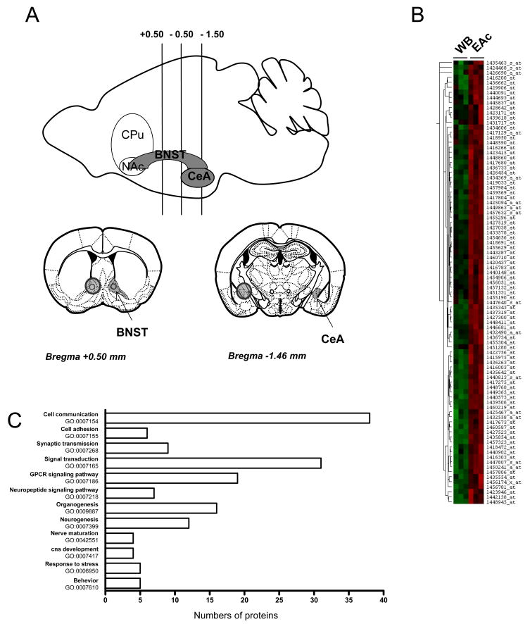
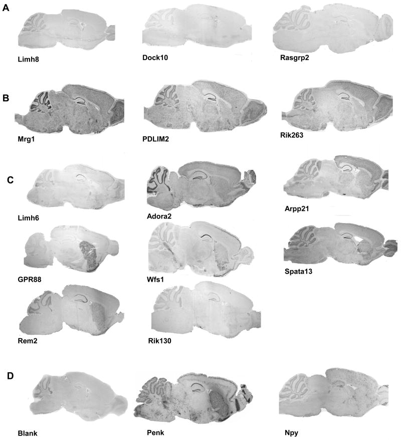
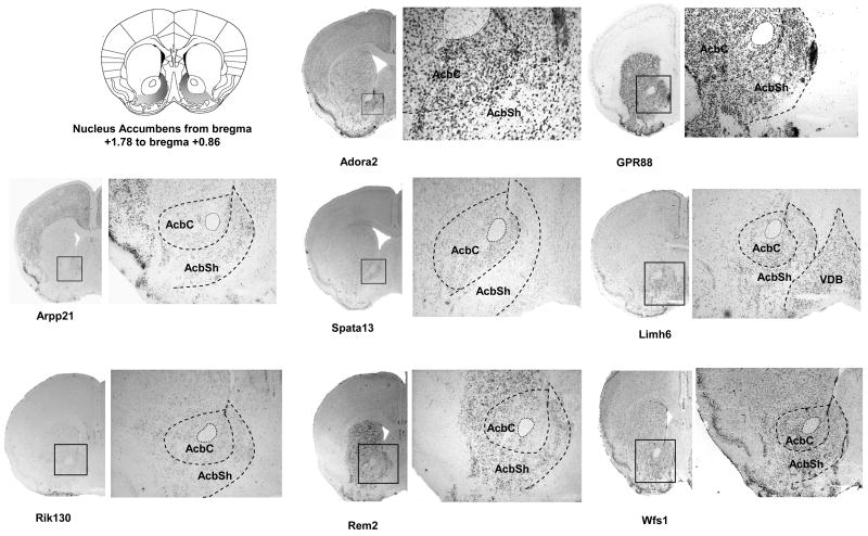
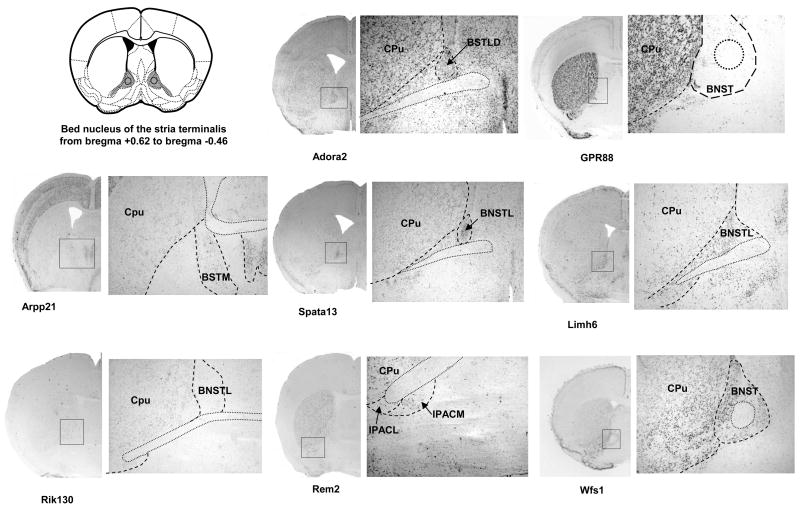
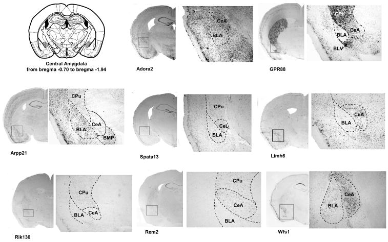
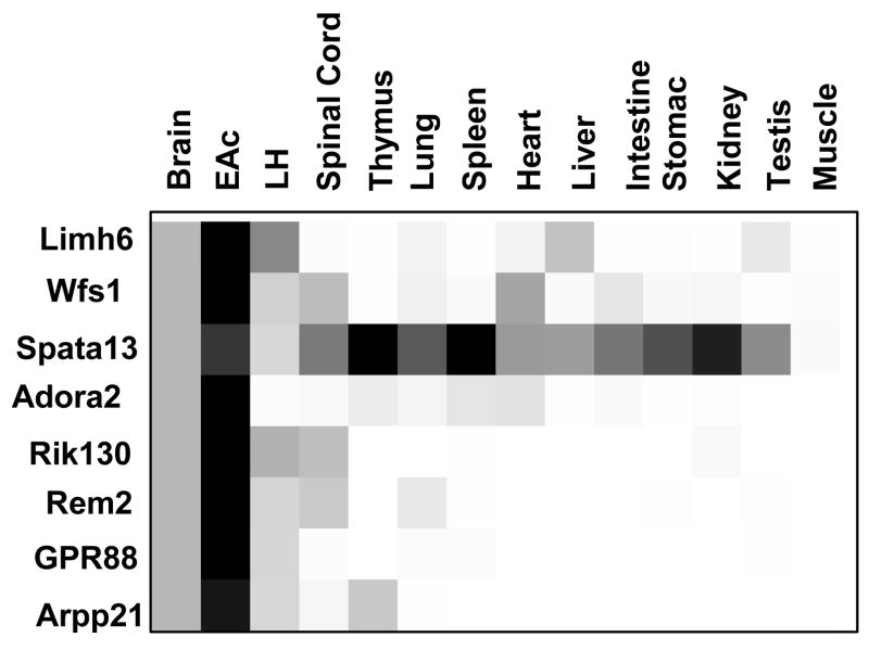
The central extended amygdala (EAc) is an ensemble of highly interconnected limbic structures of the anterior brain, and forms a cellular continuum including the bed nucleus of the stria terminalis (BNST), the central nucleus of the amygdala (CeA) and the nucleus accumbens shell (AcbSh). This neural network is a key site for interactions between brain reward and stress systems, and has been implicated in several aspects of drug abuse. In order to increase our understanding of EAc function at the molecular level, we undertook a genome-wide screen (Affymetrix) to identify genes whose expression is enriched in the mouse EAc. We focused on the less-well known BNST-CeA areas of the EAc, and identified 121 genes that exhibit more than twofold higher expression level in the EAc compared with whole brain. Among these, 43 genes have never been described to be expressed in the EAc. We mapped these genes throughout the brain, using non-radioactive in situ hybridization, and identified eight genes with a unique and distinct rostro-caudal expression pattern along AcbSh, BNST and CeA. Q-PCR analysis performed in brain and peripheral organ tissues indicated that, with the exception of one (Spata13), all these genes are predominantly expressed in brain. These genes encode signaling proteins (Adora2, GPR88, Arpp21 and Rem2), a transcription factor (Limh6) or proteins of unknown function (Rik130, Spata13 and Wfs1). The identification of genes with enriched expression expands our knowledge of EAc at a molecular level, and provides useful information to toward genetic manipulations within the EAc.


Figures

Figure 1
Figure 1. Affymetrix analysis for the identification of genes enriched in the EAc
(A) This scheme shows brain areas under study: bilateral punches (1.2 mm diameter) were taken from mouse brain coronal slices (1 mm thick) to collect the bed nucleus of stria terminalis (BNST, +0.5 to −0.5) and the central nucleus of the Amygdala (CeA, −0.5 to −1.5) (see Methods for details). BNST and CeA punches were pooled and corresponded to central Extended Amygdala (EAc) samples. (B) This hierarchical cluster illustrates raw microarray data from three independent hybridizations for the 129 selected probe sets, and shows high expression in EAc (right columns, red) compared to whole brain (WB, left columns, left). The probe set selection was based on a standard statistical analysis by MAS 5.0 and a threshold of 2-fold change in EAc over WB was used (see Methods for details). Hierarchical cluster analysis was performed using the Cluster 3.0 and Treeview softwares. (C) Gene ontology analysis of the EAc enriched genes. Genes were categorized with the Biological Process domain and significantly enriched GO terms with a probability lower than 0.01 and including at least four proteins are represented.
Figure 2
Figure 2. Expression pattern of EAc-enriched genes by in situ hybridization on sagittal brain sections
Dig-labeled RNA in situ hybridization (ISH) was performed on 25μm sagittal adult mouse brain sections 49 genes whose specific expression pattern in EAc network has not been reported earlier. In this figure, examples of low expressed genes (A), strong and ubiquitously expressed genes (B) and potential novel EAc markers (C) are shown. Neuropeptide Y (Npy) was used as a low intensity positive control, preproenkephalin (Penk) was used as a high intensity positive control and a blank hybridization was used as the negative control (D). Representative images are shown, and the complete ISH analysis on sagittal sections for all 49 genes is shown in supplemental Figure S2. Expression patterns were classified as detailed in methods. Gene symbols are indicated as in Table 1.
Figure 3
Figure 3. Expression analysis of EAc-enriched genes in AcbSh, BNST and CeA on coronal brain sections
Dig-labeled RNA in situ hybridization (ISH) was performed for a selection of 8 potential EAc markers using 25μm coronal sections of mouse brain. A scheme from the mouse brain atlas with the coordinates (Paxinos and Franklin, 2001) shows location of (A) the shell of the Nucleus Accumbens (AcbSh), (B) the bed nucleus of stria terminalis (BNST) and (C) the central nucleus of the Amygdala. Representative ISH images are shown for each candidate gene with an enlargement (zoom) for specific areas of interest. Abbreviations: AcbSh, accumbens nucleus, shell; AcbC, accumbens nucleus, core ; VDB, nucleus of the vertical limb of the diagonal band; CPu, caudate putamen; IPACL, interstitial nucleus of the posterior limb of the anterior commissure, lateral part; IPACM, interstitial nucleus of the posterior limb of the anterior commissure, medial part BSTLD, Bed Nucleus of the Stria Terminalis, lateral division, dorsal part; BSTL, Bed Nucleus of the Stria Terminalis, lateral division; BSTM, Bed Nucleus of the Stria Terminalis, medial division; BLA, basolateral amygdaloid nucleus, anterior part ; BLV, basolateral amygdaloid nucleus, ventral part CeA central amygdaloid nucleus; CeL, central amygdaloid nucleus, lateral division; BMP, basomedial amygdaloid nucleus, posterior part (Paxinos and Franklin, 2001)
Figure 4
Figure 4. Expression of EAc-enriched genes in the central nervous system and peripheral tissues by qPCR
Quantitative PCR reactions were performed in triplicate on 2 independent samples (3 mice pooled for EAc, 2 mice pooled for WB and individual n=2 mice all other tissues, see Methods for details) and data are expressed as a fold-change over WB considered as the reference sample. Each gene is represented by a single box row, with expression levels illustrated using a grey scale from white (low level) to black (high level). All the genes are mainly expressed in the central nervous system (CNS), with the exception of Spata-13 expressed in most tested tissues at levels similar or higher to CNS.

Similar articles

Cited by

References

    1. Abou-Sleymane G, Chalmel F, Helmlinger D, Lardenois A, Thibault C, Weber C, Merienne K, Mandel JL, Poch O, Devys D, Trottier Y. Polyglutamine expansion causes neurodegeneration by altering the neuronal differentiation program. Hum Mol Genet. 2006;15:691–703. - PubMed
    1. Alheid GF, Heimer L. New perspectives in basal forebrain organization of special relevance for neuropsychiatric disorders: the striatopallidal, amygdaloid, and corticopetal components of substantia innominata. Neuroscience. 1988;27:1–39. - PubMed
    1. Befort K, Filliol D, Darcq E, Ghate A, Matifas A, Lardenois A, Muller J, Thibault C, Dembele D, Poch O, Kieffer BL. Gene expression is altered in the lateral hypothalamus upon activation of the mu opioid receptor. Ann N Y Acad Sci. 2008a;1129:175–184. - PubMed
    1. Befort K, Filliol D, Ghate A, Darcq E, Matifas A, Muller J, Lardenois A, Thibault C, Dembele D, Le Merrer J, Becker JAJ, Poch O, Kieffer BL. Mu-opioid receptor activation induces transcriptional plasticity in the central extended amygdala. J neurosci. 2008b;27:2973–2984. - PubMed
    1. Bonaventure P, Guo H, Tian B, Liu X, Bittner A, Roland B, Salunga R, Ma XJ, Kamme F, Meurers B, Bakker M, Jurzak M, Leysen JE, Erlander MG. Nuclei and subnuclei gene expression profiling in mammalian brain. Brain Res. 2002;943:38–47. - PubMed

Publication types

Substances

LinkOut - more resources