. 2008 Oct 25;380(2):264-75.
doi: 10.1016/j.virol.2008.08.011. Epub 2008 Sep 10.

Kaposi's sarcoma-associated herpesvirus RTA activates the processivity factor ORF59 through interaction with RBP-Jkappa and a cis-acting RTA responsive element

Affiliations

Kaposi's sarcoma-associated herpesvirus RTA activates the processivity factor ORF59 through interaction with RBP-Jkappa and a cis-acting RTA responsive element

Yunhua Liu et al. Virology. .

Abstract

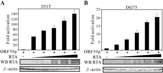
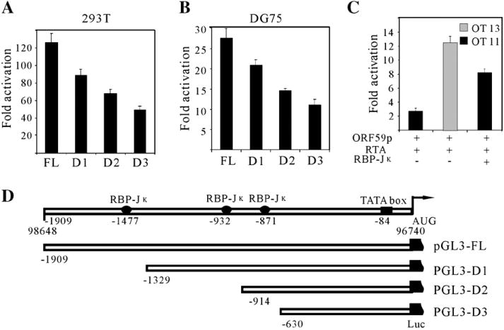
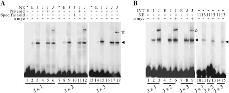
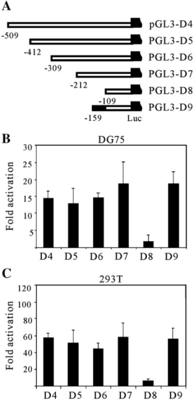
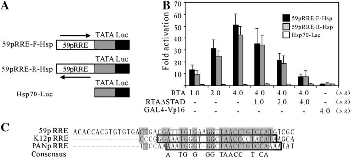
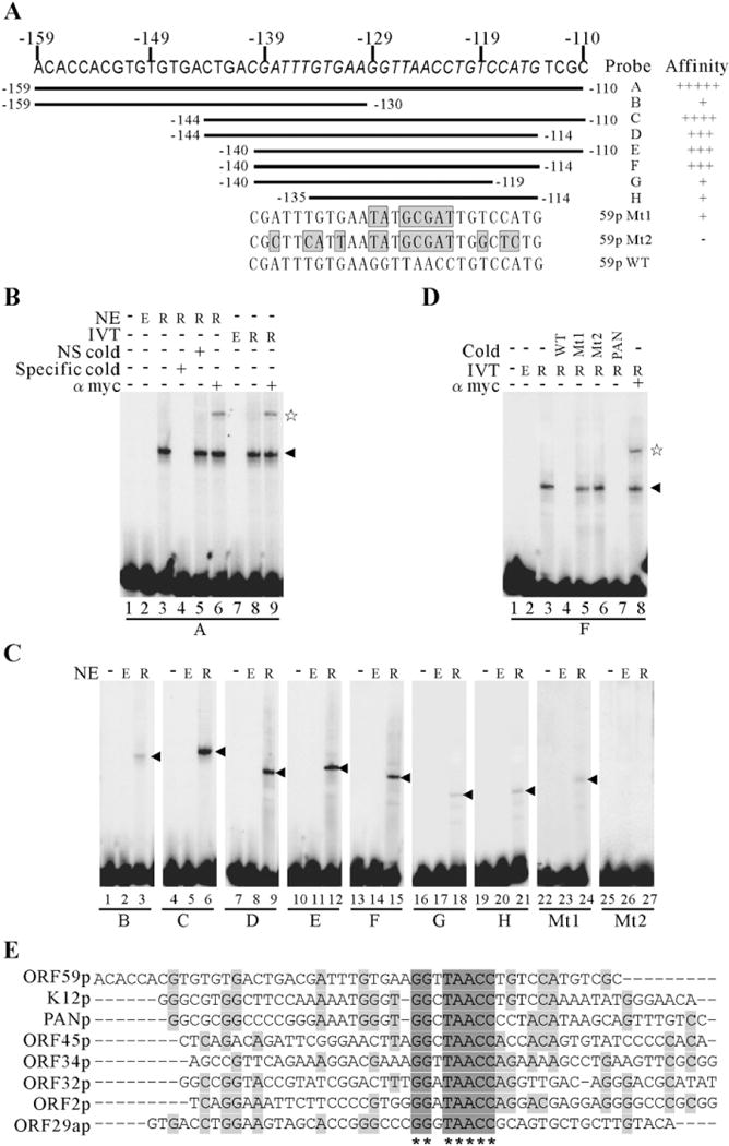
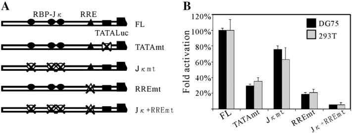
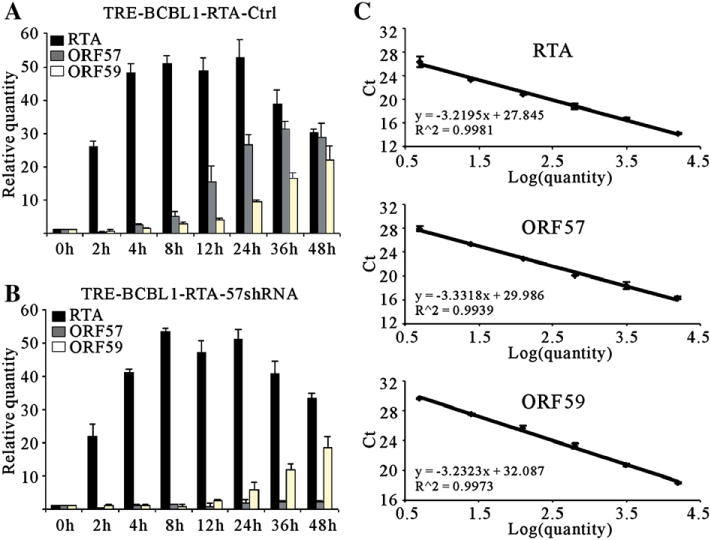
Kaposi's sarcoma-associated herpesvirus (KSHV/HHV8) displays two life modes, latency and lytic reactivation in the infected host cells which are equally important for virus mediated pathogenesis. During latency only a small number of genes are expressed. Under specific conditions, KSHV can undergo lytic replication with the production of viral progeny. One immediate-early gene RTA, encoded by open reading frame 50 of KSHV, has been shown to play a critical role in switching the viral latency to lytic reactivation. Over-expression of RTA from a heterologous promoter is sufficient for driving KSHV lytic replication which results in production of viral progeny. In the present study, we show that RTA can activate the expression of the ORF59 which encodes the processivity factor essential for DNA replication during lytic reactivation. We also show that RTA regulates ORF59 promoter through interaction with RBP-Jkappa as well as a cis-acting RTA responsive element within the promoter. In the context of KSHV infected cells, the upregulation of ORF59 is a direct response to RTA expression. Taken together, our findings provide new evidence to explain the mechanism by which RTA can regulate its downstream gene ORF59, further increasing our understanding of the biology of KSHV lytic replication.


Figures

Fig. 1
Fig. 1
Transcriptional activation of RTA on the ORF59 promoters in a dose-dependent manner. The reporter plasmid pGL3-ORF59pFL contains a 1909-bp sequence upstream of the start codon of ORF59 that drives the expression of firefly luciferase. A fixed amount (1 μg) of the reporter plasmids was cotransfected into 10 million 293T (A) and DG75 (B) cells with 0, 0.5, 1, 2.5, 5 and 10 μg of pCR3.1-RTA and 10, 9.5, 9, 7.5, 5 and 0 μg of pCR3.1 control vector. 50 ng pRL-Tk was also included in each transfection, and served as an internal control for transfection efficiency. 24 h posttransfection, cell lysate of each transfection was assayed with the dual-luciferase system. Firefly luciferase activities were normalized to the corresponding Renilla luciferase activities. Fold activation by RTA was calculated by comparing the normalized firefly luciferase activity stimulated by pCR3.1-RTA to that stimulated by pCR3.1. The means and standard deviations from three independent transfections are shown.
Fig. 2
Fig. 2
RBP-Jκ binding sites within ORF59 promoter are essential for regulation by RTA. A series of truncated promoters were cloned into a luciferase reporter plasmid pGL3-enhancer (D). Fixed amount (1 μg) of reporter plasmids were cotransfected into 293T (A) and DG75 (B) cells with either an RTA expression plasmid pCR3.1-RTA (5 μg) or pCR3.1 (5 μg). (C) ORF59 full length promoter reporter plasmid (1 μg) was transfected into a RBP-Jκ knockout cell line (OT11) and a wild type cell line (OT13) with 5 μg pCR3.1-RTA expression vector; an extra rescue experiment was carried out by introducing 5 μg of RBP-Jκ expression vector into OT11. To normalize the total amount of DNA, pcDNA 3.1 control vector was used. All the cells were harvested at 24 h posttransfection, dual-luciferase assays and fold activations were performed as described above. The means and standard deviations from three independent transfections are shown. (D) Schematic diagram of serial deletions of potential RBP-Jκ binding sites in the ORF59 promoter. Potential RBP-Jκ binding sites and TATA box are shown with the numbers referring to the nucleotide position upstream of the translation initiation site. The mapped translation initiation site is shown by the arrow.
Fig. 3
Fig. 3
RBP-Jκ binding to its cognate sequences within ORF59 promoter in vitro. (A) EMSA of the 3 putative RBP-Jκ binding sites in the ORF59 promoter with nuclear extracts (NE). Nuclear extracts (100 ng) containing RBP-Jκ protein were incubated with labeled double-stranded oligonucleotides, Jκ1 (lanes 3 to 6), Jκ2 (lanes 9 to 12) and Jκ3 (lanes 15 to 18), respectively, and the binding mixtures were then resolved on a native polyacrylamide gel. A 50-fold excess of unlabeled specific competitors (Jκ1 in lane 4; Jκ2 in lane 10 and Jκ3 in lane 16) and a nonspecific competitor (Jκ1 in lane 5; Jκ2 in lane 11 and Jκ3 in lane 17) was incubated in the presence of RBP-Jκ for the competition assay. (B) EMSA of Jκ1 (lanes 1 to 3), Jκ2 (lanes 4 to 6) and Jκ3 (lanes 7 to 9) with in vitro translated recombinant Myc-RBP-Jκ protein. End-labeled oligonucleotides were incubated with in vitro translated Myc-tagged RBP-Jκ protein (50 ng) and resolved on a native polyacrylamide gel. The DNA–protein complexes were supershifted by a monoclonal anti-Myc-tag antibody (lanes 3, 6 and 9). EMSA of Jκ1 (lanes 10, 11), Jκ2 (lanes 12, 13) and Jκ3 (lanes 14, 15) with nuclear extracts from OT11 or OT13 cells were also performed. The arrowhead denotes the specific binding of RBP-Jκ to DNA and the pentagram denotes the supershift. NE, nuclear extracts; IVT, in vitro translated; NS, nonspecific; E, empty vector; J, RBP-Jκ expression vector; 11, OT11; 13, OT13.
Fig. 4
Fig. 4
Mapping of the minimal responsive element in ORF59 promoter. (A) Scheme showing a series of truncated promoters (D4 to D9) were cloned into a luciferase reporter plasmid, pGL3-enhancer. Serial deletions were made on the 630-bp fragment from the 5′ end, with the number indicating the nucleotide position upstream of the AUG codon. Fixed amount (1 μg) of reporter plasmids were cotransfected into DG75 (B) and 293T (C) cells with either an Rta expression plasmid pCR3.1-RTA (5 μg) or pCR3.1 (5 μg). All the cells were harvested at 24 h posttransfection, dual-luciferase assays and fold activations were performed as described in Fig. 1. The means and standard deviations from three independent transfections are shown.
Fig. 5
Fig. 5
The 59pRRE confers RTA responsiveness to a heterologous promoter and possesses conservation with PAN RNA and K12 RRE. (A) 59pRRE was fused to the hsp70 TATA box (Taylor and Kingston, 1990) in forward and reverse orientations and then cloned into vector pGL3-basic. (B) The resulting plasmids, 59pRR-F-Hsp and 59pRRE-R-Hsp, and backbone alone were tested for RTA activation by reporter assays. Each was cotransfected into 293T cells with increasing amounts of ORF50 expression vector or combined with increasing amounts of the dominant negative mutant ORF50ΔSTAD as indicated. To determine the specificity of RTA activation, GAL4-VP16 was also introduced. All the cells were harvested at 24 h posttransfection, dual-luciferase assays and fold activations were performed as described in Fig. 1. The means and standard deviations from three independent transfections are shown. (C) The alignment depicts the conservation between the RREs of ORF59, PAN RNA and K12 promoter. The nucleotide sequences of three RREs were aligned by the Vector NTI Suite, version 9.0. The grey boxes denote the identical residues shared by the ORF59 promoter with either PAN RNA or K12 promoter, and the consensus is shown at the bottom. The opened boxes refer to the minimal RREs of PAN RNA and K12 promoters.
Fig. 6
Fig. 6
RTA directly binds to the 59pRRE in vitro and mapping of the core ORF59 RRE and the conservation of consensus binding sequences in other promoters. (A) Schematic representation of the oligonucleotides tested for RTA binding by EMSAs. Nucleotide residues of the 27-bp core RRE are italicized and the mutated minimal 59pRREs are shown at the bottom. The grey boxes correspond to the mutation sites. (B) EMSA of probe A with either overexpressed nuclear extracts (lanes 3 to 6) or in vitro translated recombinant Myc-RTA protein (lanes 8, 9). A 50-fold excess of unlabeled specific competitors (lane 4) and a nonspecific competitor (lane 5) was incubated in the presence of recombinant Myc-RTA protein for the competition assay. The RTA-bound complexes were supershifted by a monoclonal anti-Myc-tag antibody (lanes 6, 9). (C) Wild type oligonucleotides B to H and mutated ones were labeled and incubated in the absence or presence of nuclear extracts from empty vector or RTA expression vector transfected 293T cells, and their relative binding affinities are summarized and shown in the right of (A). (D) The specificity of the binding complex formed by 59pF and RTA. 59pF were end-labeled and incubated with in vitro translated Myc-tagged RTA protein. Competition assays were performed by inclusion 50-fold excess of unlabeled specific competitors 59pF (lane 4) or mutated 59pF (lanes 5 and 6) or PANpRRE (lane 7) in the binding reactions before incubation with 1-fold of radiolabeled probe 59pF. The specific binding complex was supershifted by a monoclonal anti-Myc-tag antibody (lane 8). The arrowhead denotes the specific binding of RTA to DNA and the pentagram denotes the supershift. NE, nuclear extracts; IVT, in vitro translated; NS, nonspecific; Mt, mutated 59pRRE; E, empty vector; R, RTA expression vector. (E) The alignment depicts the conservation of consensus binding sequences (GGNTAACC) in other KSHV promoters.
Fig. 7
Fig. 7
The contribution of TATA box, RBP-Jκ binding sites and RRE to the activation of ORF59 promoter by RTA. (A) Diagram of the mutation constructs using site-directed mutagenesis in the ORF59 promoter. All the mutated promoters are cloned into the pGL3-enhancer reporter vector and the resulting constructs are named according to the mutation sites. (B) The mutated constructs were assayed for their responsiveness to RTA in reporter assays with DG75 and 293T cell lines, as described in Fig. 2. Activity of reporter plasmids containing mutated ORF59 promoter is expressed as % activity of wild type promoter-containing reporter vector (pGL3-FL). Results are the averages of at least three experiments ±standard deviations.
Fig. 8
Fig. 8
Real-time RT-PCR analysis of RTA, ORF57 and ORF59 transcripts in TRE-BCBL1-TRE cells. The RTA-inducible BCBL-1 cells, infected with lentivirus carrying empty control pLL3.7 or ORF57 shRNA expression vectors, were treated with 5 μg/ml tetracycline and then harvested at various time points. Total RNA was extracted by TRIzol reagent. A total of 5 μg of RNA was used with the First Strand cDNA Synthesis Kit to construct cDNA. The real-time PCR was performed using the SYBR green real-time PCR master mix kit with β-actin as the standard. The relative quantitative comparison of RTA, ORF57 and ORF59 mRNA levels over the time course of induction of the TRE-BCBL1-RTA-Ctrl (A) and TRE-BCBL1-RTA-57shRNA (B) cells infected with lentivirus control or shRNA expression vectors respectively. The standard curves are shown to the right (C). Each time point was tested in triplicate for the calculation of the mean and standard deviation.

Similar articles

Cited by

References

    1. Albrecht JC, Nicholas J, Biller D, Cameron KR, Biesinger B, Newman C, Wittmann S, Craxton MA, Coleman H, Fleckenstein B, et al. Primary structure of the herpesvirus saimiri genome. J Virol. 1992;66(8):5047–5058. - PMC - PubMed
    1. Alexander L, Denekamp L, Knapp A, Auerbach MR, Damania B, Desrosiers RC. The primary sequence of rhesus monkey rhadinovirus isolate 26–95: sequence similarities to Kaposi’s sarcoma-associated herpesvirus and rhesus monkey rhadinovirus isolate 17577. J Virol. 2000;74(7):3388–3398. - PMC - PubMed
    1. AuCoin DP, Colletti KS, Cei SA, Papouskova I, Tarrant M, Pari GS. Amplification of the Kaposi’s sarcoma-associated herpesvirus/human herpesvirus 8 lytic origin of DNA replication is dependent upon a cis-acting AT-rich region and an ORF50 response element and the trans-acting factors ORF50 (K-Rta) and K8 (K-bZIP) Virology. 2004;318(2):542–555. - PubMed
    1. Buonaguro FM, Tornesello ML, Beth-Giraldo E, Hatzakis A, Mueller N, Downing R, Biryamwaho B, Sempala SD, Giraldo G. Herpesvirus-like DNA sequences detected in endemic, classic, iatrogenic and epidemic Kaposi’s sarcoma (KS) biopsies. Int J Cancer. 1996;65(1):25–28. - PubMed
    1. Bu W, Carroll KD, Palmeri D, Lukac DM. Kaposi’s sarcoma-associated herpesvirus/human herpesvirus 8 ORF50/Rta lytic switch protein functions as a tetramer. J Virol. 2007;81(11):5788–5806. - PMC - PubMed

Publication types

Substances