Review
. 2008 Dec;16(12):1433-41.
doi: 10.1016/j.joca.2008.06.016. Epub 2008 Sep 10.

The osteoarthritis initiative: report on the design rationale for the magnetic resonance imaging protocol for the knee

Affiliations
Review

The osteoarthritis initiative: report on the design rationale for the magnetic resonance imaging protocol for the knee

C G Peterfy et al. Osteoarthritis Cartilage. 2008 Dec.

Abstract

Objectives: To report on the process and criteria for selecting acquisition protocols to include in the osteoarthritis initiative (OAI) magnetic resonance imaging (MRI) study protocol for the knee.

Methods: Candidate knee MR acquisition protocols identified from the literature were first optimized at 3Tesla (T). Twelve knees from 10 subjects were scanned one time with each of 16 acquisitions considered most likely to achieve the study goals and having the best optimization results. The resultant images and multi-planar reformats were evaluated for artifacts and structural discrimination of articular cartilage at the cartilage-fluid, cartilage-fat, cartilage-capsule, cartilage-meniscus and cartilage-cartilage interfaces.

Results: The five acquisitions comprising the final OAI MRI protocol were assembled based on the study goals for the imaging protocol, the image evaluation results and the need to image both knees within a 75 min time slot, including positioning. For quantitative cartilage morphometry, fat-suppressed, 3D dual-echo in steady state (DESS) acquisitions appear to provide the best universal cartilage discrimination.

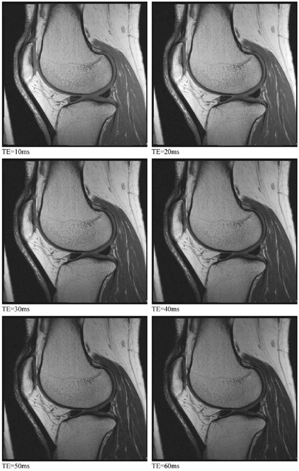
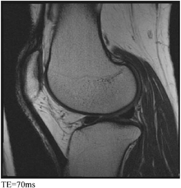
Conclusions: The OAI knee MRI protocol provides imaging data on multiple articular structures and features relevant to knee OA that will support a broad range of existing and anticipated measurement methods while balancing requirements for high image quality and consistency against the practical considerations of a large multi-center cohort study. Strengths of the final knee MRI protocol include cartilage quantification capabilities in three planes due to multi-planar reconstruction of a thin slice, high spatial resolution 3D DESS acquisition and the multiple, non-fat-suppressed image contrasts measured during the T2 relaxation time mapping acquisition.


Conflict of interest statement

Conflict of interest None of the authors have any financial or other interests related to the manuscript submitted to Osteoarthritis and Cartilage that might constitute a potential conflict of interest.

Figures

Fig. 1
Fig. 1
Orientation of coronal acquisitions. Coronal 2D and 3D acquisitions are prescribed coronal to the joint, with the slice axis parallel to the long axis of the femoral diaphysis on the sagittal localizer (A) and to a line tangent to the posterior cortices of the femoral condyles on the axial localizer (B). Depiction of both posterior femoral cortices (arrows) within two slices (3 mm) of each other confirms proper alignment on this example of COR 3D FLASH WE (C).
Fig. 2
Fig. 2
Example of COR IW 2D TSE. Note delineation of the MCL, LCL, body segments of the menisci, central tibial and femoral bone margins and the central tibiofemoral articular cartilage. Note that chemical-shift artifact is relatively mild.
Fig. 3
Fig. 3
Orientation of the sagittal acquisitions. Anatomical coverage on sagittal 2D and 3D acquisitions should include the tibial tubercle, the entire patella and as much of the suprapatellar bursa as possible (A) Sagittal acquisitions are prescribed orthogonal to the coronal acquisitions and sagittal to the joint, with the slice axis parallel to the long axis of the femoral diaphysis on the coronal localizer (B) and perpendicular to a line tangent to the posterior cortices of the femoral condyles on the axial localizer (C).
Fig. 4
Fig. 4
SAG 3D DESS WE. Note the clear delineation of the cartilage-cartilage (small arrows) and cartilage-capsule (large arrow) interfaces as well as the interfaces between cartilage and adipose (F), bone (B) and meniscus (M).
Fig. 5
Fig. 5
Coronal MPR of SAG 3D DESS WE. Orientation is identical to that described in Fig. 1. Note the excellent delineation of the cartilage–fluid interface (arrow), and the high contrast between cartilage and bone (B) and cartilage and meniscus (M).
Fig. 6
Fig. 6
Axial MPR of SAG 3D DESS WE. Axial coverage includes any superior or inferior patellar osteophytes and extends to the tibial epiphysis. Note the good cartilage–fluid contrast revealing thinning of articular cartilage over the lateral facet of the patella (large arrow). Note also, that a small aliasing artifact (small arrow) is present at the top of the image but does not obscure any anatomy of interest.
Fig. 7
Fig. 7
Sensitivity to subarticular BMA and cysts. SAG IW 2D TSE FS (A) shows both bone cysts (small arrow) and surrounding BMA (large arrow) in the femoral trochlea of this knee. However, both GRE scans, SAG 3D DESS WE (B) and COR 3D FLASH WE (C), of the same knee show only the cysts in this location.
Fig. 8
Fig. 8
SAG 2D MESE. Multiple contrast acquisitions having progressively longer TEs can be combined to generate T2 maps of the articular cartilage and adjacent tissues. These seven images illustrate how changing the TE affects the relative signal and relative contrast among the different tissues in the knee.
Fig. 8
Fig. 8
SAG 2D MESE. Multiple contrast acquisitions having progressively longer TEs can be combined to generate T2 maps of the articular cartilage and adjacent tissues. These seven images illustrate how changing the TE affects the relative signal and relative contrast among the different tissues in the knee.

Similar articles

Cited by

References

    1. Peterfy CG, Gold G, Eckstein F, Cicuttini F, Dardzinski B, Stevens R. MRI protocols for whole-organ assessment of the knee in osteoarthritis. Osteoarthritis Cartilage. 2006;14(Suppl A):A95–A111. - PubMed
    1. Gold GE, Burstein D, Dardzinski B, Lang P, Boada F, Mosher T. MRI of articular cartilage in OA: novel pulse sequences and compositional/-functional markers. Osteoarthritis Cartilage. 2006;14(Suppl A):A76–86. - PubMed
    1. Eckstein F, Cicuttini F, Raynauld JP, Waterton JC, Peterfy C. Magnetic resonance imaging (MRI) of articular cartilage in knee osteoarthritis (OA): morphological assessment. Osteoarthritis Cartilage. 2006;14(Suppl A):A46–75. - PubMed
    1. Eckstein F, Mosher T, Hunter D. Imaging of knee osteoarthritis: data beyond the beauty. Curr Opin Rheumatol. 2007;19(5):435–43. - PubMed
    1. Burstein D, Gray ML. Is MRI fulfilling its promise for molecular imaging of cartilage in arthritis? Osteoarthritis Cartilage. 2006;14(11):1087–90. - PubMed

Publication types