. 2008 Nov 14;283(46):31371-7.
doi: 10.1074/jbc.M802814200. Epub 2008 Sep 11.

Prointerleukin-18 is activated by meprin beta in vitro and in vivo in intestinal inflammation

Affiliations

Prointerleukin-18 is activated by meprin beta in vitro and in vivo in intestinal inflammation

Sanjita Banerjee et al. J Biol Chem. .

Abstract

Interleukin-18 (IL-18), a pro-inflammatory cytokine, is a key factor in inflammatory bowel disease (IBD). Caspase-1 activates this cytokine, but other proteases are likely involved in maturation. Because meprin metalloproteinases have been implicated in IBD, the interaction of these proteases with proIL-18 was studied. The results demonstrate that the meprin beta subunit of meprins A and B cleaves proIL-18 into a smaller 17-kDa product. The cleavage is at the Asn51-Asp52 bond, a site C-terminal to caspase-1 cleavage. The cleavage occurred in vitro with a Km of 1.3 microm and in Madin-Darby canine kidney cells transfected with meprin beta when proIL-18 was added to the culture medium. The product of meprin B cleavage of proIL-18 activated NF-kappaB in EL-4 cells, indicating that it was biologically active. To determine the physiological significance of the interactions of meprins with proIL-18, an experimental model of IBD was produced by administering dextran sulfate sodium (DSS) to wild-type and meprin beta knock-out (betaKO) mice, and the serum levels of active IL-18 were determined. DSS-treated meprin betaKO mice had lower levels of the active cytokine in the serum compared with wild-type mice. Furthermore, in meprin alphaKO mice, which express meprin beta but not alpha, active IL-18 was elevated in the serum of DSS-treated mice compared with wild-type mice, indicating that the meprin isoforms have opposing effects on the IL-18 levels in vivo. This study identifies proIL-18 as a biologically important substrate for meprin beta and implicates meprins in the modulation of inflammation.


Figures

FIGURE 1.
FIGURE 1.
Expression and purification of proIL-18. A, vector map of pET28a::IL-18. ProIL-18 was cloned at the NdeI-EcoRI sites. The N-terminal 6-histidine tag and thrombin cleavage site are indicated. B, Coomassie Blue staining of Ni2+ purification of His6-proIL-18 after isopropyl β-d-thiogalactopyranoside induction. Lane 1, sample added to Ni2+ column; lane 2, flow-through fraction; lanes 3–14, eluate fractions. C, silver staining of purified proIL-18 before and after thrombin cleavage. Lane 1, His6-proIL-18; lane 2, thrombin-cleaved proIL-18; lanes 3–8, eluate fractions after Ni2+ purification.
FIGURE 2.
FIGURE 2.
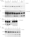
Cleavage of proIL-18 by meprin isoforms. A and B, proIL-18 (2.2 μm) was incubated with meprin isoforms (0.11 μm) as described under “Experimental Procedures.” The mixtures were subjected to SDS-PAGE, and the substrates and products were visualized by silver staining (A) or by immunoblotting using a polyclonal antibody to IL-18 (B). Meprin isoforms were homomeric meprin A (lanes 1–3), heteromeric meprin A (lanes 4–6), and meprin B (lanes 7–9). C, the hydrolysis of proIL-18 by meprin B was inhibited when actinonin (35 μm) was included in the incubation mixture. D, proIL-18 (2.2 μm) was incubated with 2 nm meprin B for up to 12 h, and the proteins were visualized by silver staining.
FIGURE 3.
FIGURE 3.
Identification of the proIL-18 site cleaved by meprin B. The meprin B cleavage site is indicated, and the nine-amino acid sequence identified by MS/MS is shown in boldface. Caspase-1 and putative PR-3 sites are also indicated. The region of caspase-3 cleavage is italicized (1).
FIGURE 4.
FIGURE 4.
Interaction of proIL-18 with meprin B expressed in MDCK cells. Full-length meprin β was transfected into MDCK cells, and latent meprin B was activated with trypsin. After removal and inhibition of trypsin, proIL-18 was added to the culture medium and incubated for 22 h; the medium and the cell lysate were then subjected to SDS-PAGE. The upper panel is a Western blot of the culture medium using a polyclonal antibody to IL-18; the lower panel is a Western blot of the cell lysate fraction using a polyclonal antibody to meprin β. The results are typical of three independent experiments.
FIGURE 5.
FIGURE 5.
Effect of meprin B on IL-18 activity in EL-4 cells. NF-κB activation was used to assess IL-18 bioactivity. EL-4 cells were untreated or treated for 30 min with proIL-18 (200 ng/ml) alone, meprin B (mepB) alone, or proIL-18 preincubated with meprin B. Cells lysates were assayed for NF-κB activation using a p65 TransAM kit. Jurkat nuclear extract served as a positive control for NF-κB activation. NF-κB activation is shown as -fold increase over unstimulated EL-4 cells (n = 3; *, p < 0.02). Results are presented as means of three independent experiments.
FIGURE 6.
FIGURE 6.
Measurement of active IL-18 levels in the serum of DSS-treated meprinβKO andαKO mice and their corresponding WT littermates. IBD was induced by administering 3.5% DSS in the drinking water for 4 days and water on the 5th day (9). Controls (Con) were given water. Serum was collected on days 3, 4, and 5, and the levels of active IL-18 were measured by enzyme-linked immunosorbent assay. A,WTand βKO mice had increased levels of active IL-18 compared with their respective control groups (n = five/group; *, p < 0.02, DSS-treated WT versus control; #, p < 0.0002, DSS-treatedβKO versus control). Meprin βKO mice treated with DSS showed significantly lower levels of serum IL-18 compared with WT mice given DSS treatment (**, p < 0.05). B, meprin αKO mice showed significantly elevated levels of IL-18 compared with WT mice after the same DSS treatment (n = seven/group; **, p < 0.0015). Both DSS-treated groups showed significant elevation in their serum IL-18 levels compared with the respective control populations (*, p < 0.0002, DSS-treated WT versus control; #, p < 5 × 10–11, DSS-treated αKO versus control).

Similar articles

Cited by

References

    1. Fantuzzi, G., and Dinarello, C. A. (1999) J. Clin. Immunol. 19 1–11 - PubMed
    1. Reuter, B. K., and Pizarro, T. T. (2004) Eur. J. Immunol. 34 2347–2355 - PubMed
    1. Mehta, V. B., Hart, J., and Wewers, M. D. (2001) J. Biol. Chem. 276 3820–3826 - PubMed
    1. Andrei, C., Margiocco, P., Poggi, A., Lotti, L. V., Torrisi, M. R., and Rubartelli, A. (2004) Proc. Natl. Acad. Sci. U. S. A. 101 9745–9750 - PMC - PubMed
    1. Bond, J. S., Matters, G. L., Banerjee, S., and Dusheck, R. E. (2005) FEBS Lett. 579 3317–3322 - PubMed

Publication types

MeSH terms