

Adventitial transplantation of blood outgrowth endothelial cells in porcine haemodialysis grafts alleviates hypoxia and decreases neointimal proliferation through a matrix metalloproteinase-9-mediated pathway--a pilot study
- PMID: 18786975
- PMCID: PMC2639314
- DOI: 10.1093/ndt/gfn433
Adventitial transplantation of blood outgrowth endothelial cells in porcine haemodialysis grafts alleviates hypoxia and decreases neointimal proliferation through a matrix metalloproteinase-9-mediated pathway--a pilot study
Abstract
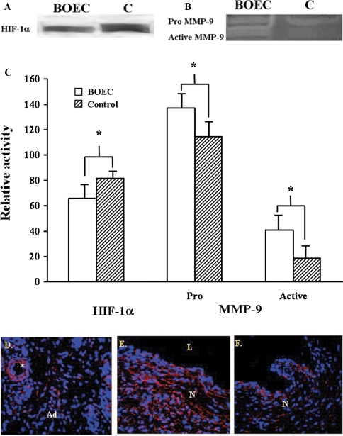
Purpose. We hypothesized that adventitial transplantation of blood outgrowth endothelial cells (BOEC) to the vein-to-graft anastomosis of polytetrafluoroethylene grafts will reduce neointimal hyperplasia by reducing hypoxia inducible factor-1alpha (HIF-1alpha), by increasing angiogenesis in a porcine model of chronic renal insufficiency with haemodialysis polytetrafluoroethylene grafts. Because matrix metalloproteinases (MMPs) have been shown to be involved with angiogenesis, the expression of MMPs and their inhibitors was determined.
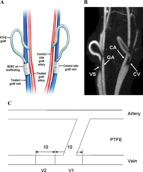
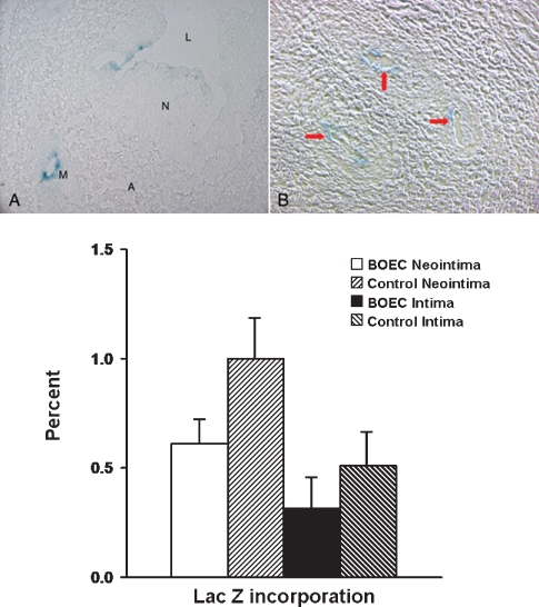
Methods: Chronic renal insufficiency was created by subtotal renal infarction and 28 days later, arteriovenous PTFE grafts were placed bilaterally from the carotid artery to the jugular vein. Autologous blood outgrowth endothelial cells labeled with Lac Z were transplanted to the adventitia of the vein-to-graft anastomosis using polyglycolic acid scaffolding and scaffolding only to other side (control). Animals were killed 14 days later and vessels were explanted from the vein-to-graft anastomosis of both sides and underwent immunohistochemical analysis, western blotting and zymography for HIF-1alpha, MMP-2, MMP-9, TIMP-1 and TIMP-2. BOEC were also made hypoxic and normoxic for 12, 24 and 48 h to determine protein expression for MMPs and TIMPs.
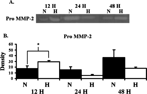
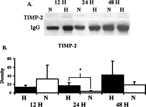
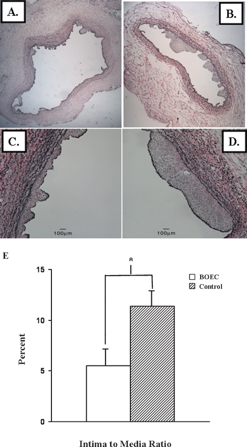
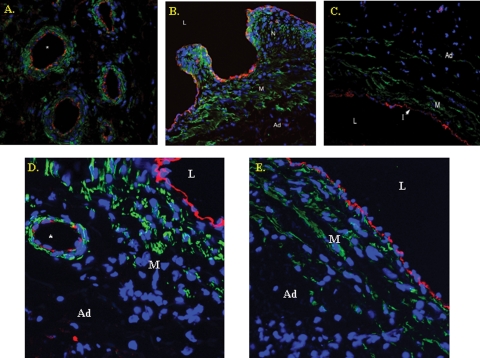
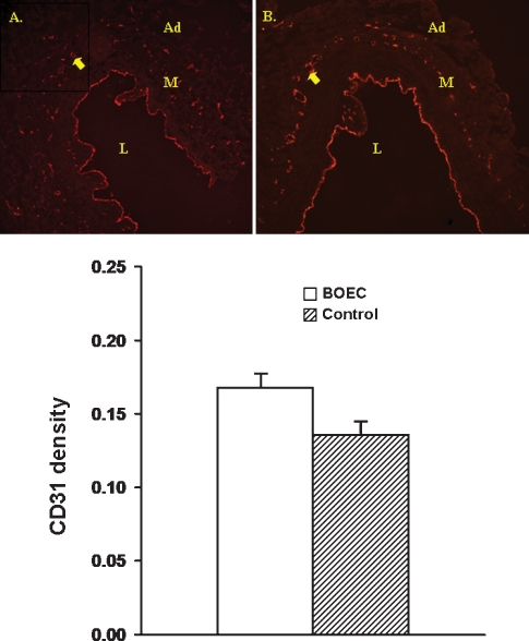
Results: Under hypoxia, BOEC significantly increased the expression of pro MMP-2 by 12 h and TIMP-2 by 24 h when compared to normoxic cells (P < 0.05). Transplantation of BOEC resulted in a significant decrease in both HIF-1alpha and intima-to-media ratio with a significant increase in both pro and active MMP-9 when compared to control vessels (P < 0.05). MMP-9 activity was localized to the neointima of the transplanted vessels by immunohistochemistry. There was increased CD31 density with engraftment of BOEC cells into the neointima of both the transplanted vessels compared to controls (P = NS).
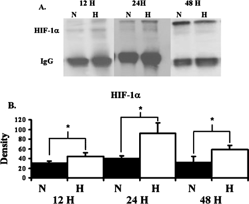
Conclusion: Transplantation of BOEC resulted in a significant decrease in intimal hyperplasia and HIF-1alpha with a significant increase in both pro and active MMP-9 that was localized to the neointima of transplanted vessels. The increase in MMP-9 offers a possible mechanism for angiogenesis and the reduced intima-to-media ratio. Furthermore, we observed that BOEC had homed to the neointima of the contralateral vessels that had increased levels of HIF-1alpha, suggesting that hypoxia may be an important stimulus for BOEC migration.
Figures
Similar articles
-
Hypoxia-induced phenotypic switch of fibroblasts to myofibroblasts through a matrix metalloproteinase 2/tissue inhibitor of metalloproteinase-mediated pathway: implications for venous neointimal hyperplasia in hemodialysis access.J Vasc Interv Radiol. 2010 Jun;21(6):896-902. doi: 10.1016/j.jvir.2010.02.030. J Vasc Interv Radiol. 2010. PMID: 20434368 Free PMC article.
-
Expression of hypoxia inducible factor-1 alpha, macrophage migration inhibition factor, matrix metalloproteinase-2 and -9, and their inhibitors in hemodialysis grafts and arteriovenous fistulas.J Vasc Interv Radiol. 2008 Feb;19(2 Pt 1):252-9. doi: 10.1016/j.jvir.2007.10.031. J Vasc Interv Radiol. 2008. PMID: 18341958
-
Increased shear stress with upregulation of VEGF-A and its receptors and MMP-2, MMP-9, and TIMP-1 in venous stenosis of hemodialysis grafts.Am J Physiol Heart Circ Physiol. 2008 May;294(5):H2219-30. doi: 10.1152/ajpheart.00650.2007. Epub 2008 Mar 7. Am J Physiol Heart Circ Physiol. 2008. PMID: 18326810 Free PMC article.
-
Differential expression and activity of matrix metalloproteinases during flow-modulated vein graft remodeling.J Vasc Surg. 2004 May;39(5):1084-90. doi: 10.1016/j.jvs.2003.12.031. J Vasc Surg. 2004. PMID: 15111865
-
Pressure distention compared with pharmacologic relaxation in vein grafting upregulates matrix metalloproteinase-2 and -9.J Vasc Surg. 2005 Oct;42(4):747-56. doi: 10.1016/j.jvs.2005.05.037. J Vasc Surg. 2005. PMID: 16242564
Cited by
-
Hemodynamic Shear Stress and Endothelial Dysfunction in Hemodialysis Access.Open Urol Nephrol J. 2014;7(Suppl 1 M5):33-44. doi: 10.2174/1874303X01407010033. Open Urol Nephrol J. 2014. PMID: 25309636 Free PMC article.
-
The molecular mechanisms of hemodialysis vascular access failure.Kidney Int. 2016 Feb;89(2):303-316. doi: 10.1016/j.kint.2015.12.019. Kidney Int. 2016. PMID: 26806833 Free PMC article. Review.
-
Pleotropic effects of hypoxia-inducible factor-prolyl hydroxylase domain inhibitors: are they clinically relevant?Kidney Res Clin Pract. 2023 Jan;42(1):27-38. doi: 10.23876/j.krcp.22.118. Epub 2022 Nov 21. Kidney Res Clin Pract. 2023. PMID: 36634968 Free PMC article.
-
Hypoxia-induced phenotypic switch of fibroblasts to myofibroblasts through a matrix metalloproteinase 2/tissue inhibitor of metalloproteinase-mediated pathway: implications for venous neointimal hyperplasia in hemodialysis access.J Vasc Interv Radiol. 2010 Jun;21(6):896-902. doi: 10.1016/j.jvir.2010.02.030. J Vasc Interv Radiol. 2010. PMID: 20434368 Free PMC article.
-
Disruptive technological advances in vascular access for dialysis: an overview.Pediatr Nephrol. 2018 Dec;33(12):2221-2226. doi: 10.1007/s00467-017-3853-7. Epub 2017 Nov 29. Pediatr Nephrol. 2018. PMID: 29188361 Review.
References
-
- Collins AJ, Kasiske B, Herzog C, et al. Excerpts from the United States renal data system 2003 annual data report: atlas of end-stage renal disease in the United States. Am J Kidney Dis. 2003;42(Suppl 5):A5–A7. - PubMed
-
- Schwab SJ, Harrington JT, Singh A, et al. Vascular access for haemodialysis. Kidney Int. 1999;55:2078–2090. - PubMed
-
- Sullivan KL, Besarab A, Bonn J, et al. Haemodynamics of failing dialysis grafts. Radiology. 1993;186:867–872. - PubMed
-
- Roy-Chaudhury P, Kelly BS, Miller MA, et al. Venous neointimal hyperplasia in polytetrafluoroethylene dialysis grafts. Kidney Int. 2001;59:2325–2334. - PubMed
-
- Semenza GL. Targeting HIF-1 for cancer therapy. Nat Rev Cancer. 2003;3:721–732. - PubMed
Publication types
MeSH terms
Substances
Grants and funding
LinkOut - more resources
Full Text Sources
Medical
Research Materials
Miscellaneous
