. 2008 Nov;82(22):10964-74.
doi: 10.1128/JVI.01646-08. Epub 2008 Sep 10.

Early B-cell activation after West Nile virus infection requires alpha/beta interferon but not antigen receptor signaling

Affiliations

Early B-cell activation after West Nile virus infection requires alpha/beta interferon but not antigen receptor signaling

Whitney E Purtha et al. J Virol. 2008 Nov.

Abstract

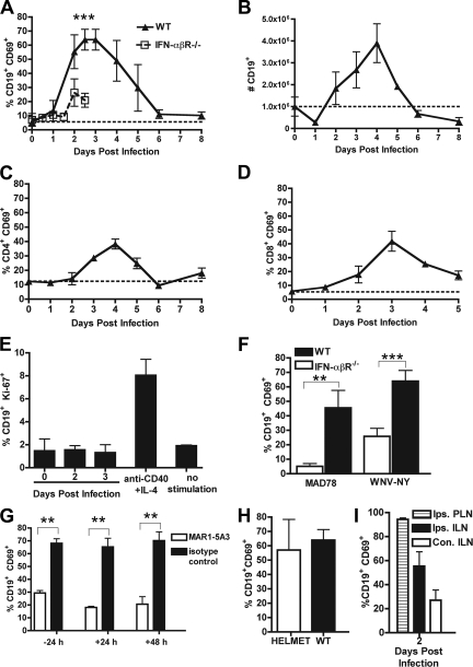
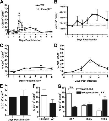
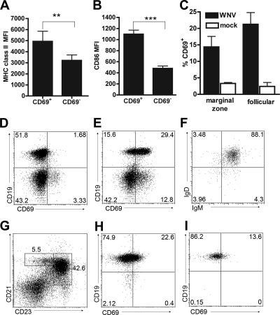
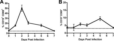
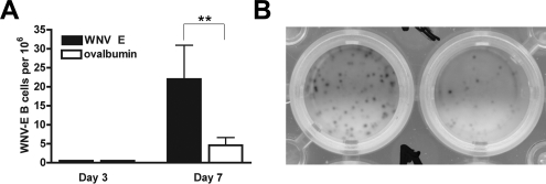
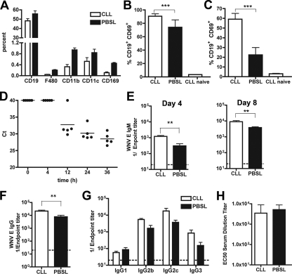
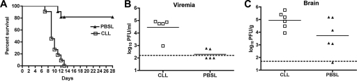
The B-cell response against West Nile virus (WNV), an encephalitic Flavivirus of global concern, is critical to controlling central nervous system dissemination and neurological sequelae, including death. Here, using a well-characterized mouse model of WNV infection, we examine the factors that govern early B-cell activation. Subcutaneous inoculation with a low dose of replicating WNV results in extensive B-cell activation in the draining lymph node (LN) within days of infection as judged by upregulation of the surface markers CD69, class II major histocompatibility complex, and CD86 on CD19(+) cells. B-cell activation in the LN but not the spleen was dependent on signals through the type I alpha/beta interferon (IFN-alpha/beta) receptor. Despite significant activation in the draining LN at day 3 after infection, WNV-specific B cells were not detected by immunoglobulin M enzyme-linked immunospot analysis until day 7. Liposome depletion experiments demonstrate that B-cell activation after WNV infection was not affected by the loss of F4/80(+) or CD169(+) subcapsular macrophages. Nonetheless, LN myeloid cells were essential for control of viral replication and survival from infection. Overall, our data suggest that the massive, early polyclonal B-cell activation occurring in the draining LN after WNV infection is immunoglobulin receptor and macrophage independent but requires sustained signals through the type I IFN-alpha/beta receptor.


Figures

FIG. 1.
FIG. 1.
B-cell activation in the draining LN. A. Expression of CD69 on CD19+ B cells after WNV-NY infection in wild-type (WT) and IFN-α/βR−/− mice. The data reflect averages from 5 to 10 mice per time point. B. Total number of CD69+ CD19+ B cells in the ILN after WNV-NY infection. C and D. Expression of CD69 on CD4+ (C) and CD8+ (D) T cells after WNV-NY infection. E. Measurement of B-cell proliferation in ILN at the indicated days after WNV infection or mitogenic stimulus (anti-CD40 plus IL-4) as judged by Ki-67 expression. F. Expression of CD69 on CD19+ B cells in wild-type and IFN-α/βR−/− mice 3 days after infection with WNV-MAD or WNV-NY. G. Effect of neutralizing anti-IFN-α/βR MAb (MAR1-5A3) on CD69 expression on CD19+ B cells after WNV infection. Mice were treated with MAb (or isotype control) at the indicated times relative to infection, and B cells were analyzed at 3 days after infection. H. Expression of CD69 on CD19+ B cells in HELMET mice 3 days after WNV infection. The dotted line represents the basal level of expression in naïve wild-type mice. Asterisks indicate time points at which differences are statistically significant (**, P < 0.01; ***, P < 0.001). I. Expression of CD69 on CD19+ B cells from the ipsilateral popliteal lymph node (PLN), ipsilateral ILN, and contralateral ILN node 2 days after WNV infection.
FIG. 2.
FIG. 2.
B-cell activation in the spleen. A. Expression of CD69 on CD19+ splenic B cells after WNV-NY infection in wild-type and IFN-α/βR−/− mice. The data reflect averages from 5 to 10 mice per time point. B. Total number of CD69+ CD19+ B cells in the spleen after WNV-NY infection. C and D. Expression of CD69 on CD4+ (C) and CD8+ (D) T cells after WNV-NY infection. E. Measurement of B-cell proliferation in the spleen at the indicated days before or after WNV infection as judged by Ki-67 expression. F. Expression of CD69 on CD19+ B cells in HELMET mice 3 days after WNV infection. G. Effect of neutralizing anti-IFN-α/βR MAb (MAR1-5A3) on CD69 expression on splenic B cells after WNV-NY infection. Mice were treated with MAb (or isotype control) at the indicated times relative to infection, and B cells were analyzed at 3 days after infection. The dotted line represents the basal level of expression in naïve wild-type mice. Asterisks indicate time points at which differences are statistically significant (*, P < 0.05; **, P < 0.01).
FIG. 3.
FIG. 3.
Phenotyping of activated B cells in LN and spleen after WNV infection. A and B. B-cell activation in the draining LN at day 3 after WNV infection was assessed by immunostaining for increased surface expression of MHC class II (A) and CD86 (B). Three-color staining was performed with CD19+ CD69+ and CD19+ CD69 cells. Asterisks indicate differences that are statistically significant (**, P < 0.01; ***, P < 0.001). D and E. WNV infection induces CD69 expression on CD19+ B cells. A representative flow cytometry panel of LN cells from uninfected (D) and WNV-infected (E) mice is shown. F. All of the CD69+ B cells in the LN showed a mature (surface IgD+ IgM+) phenotype. G to I. CD69 staining on different B-cell subsets in the spleen at day 4 after WNV infection. (H and I) Follicular (CD21high CD23high) (H) and marginal zone (CD21high CD23low) (I) B cells were determined by flow cytometry after gating (G). The average values for activation of these B-cell subsets are indicated in panel C and reflect results for at least five mice per condition.
FIG. 4.
FIG. 4.
B-cell activation after MNV infection. CD69 expression on CD19+ B cells from the draining mesenteric lymph node (A) or spleen (B) after peroral infection with MNV. Each time point represents the average of at least five mice.
FIG. 5.
FIG. 5.
Antigen-specific IgM ELISPOT in the ILN after WNV infection. A. Three or 7 days after infection with WNV, cells from the ILN were harvested and analyzed using an IgM ELISPOT against specific (WNV E protein) or nonspecific (ovalbumin) protein. The data are expressed as the number of antigen-specific B cells producing IgM per 106 cells and are averages of at least six mice per time point. B. A digital photographic example of an ELISPOT from B cells harvested at day 7 after WNV infection. The left well represents B cells that recognize WNV-E, and the right well represents nonspecific B cells that bind ovalbumin.
FIG. 6.
FIG. 6.
Effects of LN macrophage depletion on B-cell activation and humoral response after WNV infection. Mice were treated with clodronate- or PBS-containing liposomes (CLL or PBSL, respectively) 1 week prior to infection with WNV-NY. A. Evidence of depletion of F4/80+ and CD169+ macrophages in the ILN by clodronate liposomes immediately prior to infection. Samples were processed by multicolor flow cytometry. Depletion with clodronate-containing liposomes reduced the number of F4/80+ and CD169+ macrophages by 78 and 76%, respectively. B and C. Effect of clodronate or PBS liposomes on B-cell activation as judged by CD69 expression after WNV infection in the ILN (B) and spleen (C). Cells were processed 3 (ILN) or 4 (spleen) days after infection. D. Time course of appearance of WNV RNA in the draining LN as judged by fluorogenic quantitative reverse transcription-PCR. The data are expressed as Ct values, where a value of 40 is considered negative. E and F. Effect of clodronate on WNV-specific IgM (E) and IgG (F) titers at days 4 and 8 after infection. Data are expressed as reciprocal log endpoint titers after regression analysis and reflect at least eight mice per group. G. IgG subclass analysis after clodronate- or PBS-liposome treatment. Samples were from day 8 after infection. No statistically significant differences were observed. H. Relative neutralizing activity of serum from mice treated with clodronate or PBS liposomes at day 8 after WNV infection. Samples were analyzed using a previously described reporter virus particle assay on Raji-DC-SIGN-R cells (40). Data are expressed as the EC50 titer (concentration at which 50% neutralization occurs) and reflect results for five mice per group. Asterisks represent statistically significant differences (**, P < 0.01; ***, P < 0.001).
FIG. 7.
FIG. 7.
Effects of LN macrophage depletion on WNV pathogenesis. Mice were treated with clodronate- or PBS-containing liposomes (CLL or PBSL, respectively) as described above and then infected with 102 PFU of WNV-NY. Animals were monitored for survival (n = 11 per group; P < 0.0001) (A) or viral burden as measured by plaque assay from serum at day 3 (B) or brain at day 8 (C) after infection. The dotted lines indicate the limit of sensitivity of the assay.

Similar articles

Cited by

References

    1. Adachi, O., T. Kawai, K. Takeda, M. Matsumoto, H. Tsutsui, M. Sakagami, K. Nakanishi, and S. Akira. 1998. Targeted disruption of the MyD88 gene results in loss of IL-1- and IL-18-mediated function. Immunity 9143-150. - PubMed
    1. Alexopoulou, L., A. C. Holt, R. Medzhitov, and R. A. Flavell. 2001. Recognition of double-stranded RNA and activation of NF-κB by Toll-like receptor 3. Nature 413732-738. - PubMed
    1. Alsharifi, M., M. Lobigs, M. Regner, E. Lee, A. Koskinen, and A. Mullbacher. 2005. Type I interferons trigger systemic, partial lymphocyte activation in response to viral infection. J. Immunol. 1754635-4640. - PubMed
    1. Ben-Nathan, D., I. Huitinga, S. Lustig, N. van Rooijen, and D. Kobiler. 1996. West Nile virus neuroinvasion and encephalitis induced by macrophage depletion in mice. Arch. Virol. 141459-469. - PubMed
    1. Bernasconi, N. L., E. Traggiai, and A. Lanzavecchia. 2002. Maintenance of serological memory by polyclonal activation of human memory B cells. Science 2982199-2202. - PubMed

Publication types

MeSH terms