. 2008 Nov;82(22):11152-66.
doi: 10.1128/JVI.01519-08. Epub 2008 Sep 10.

Multiple virus lineages sharing recent common ancestry were associated with a Large Rift Valley fever outbreak among livestock in Kenya during 2006-2007

Affiliations

Multiple virus lineages sharing recent common ancestry were associated with a Large Rift Valley fever outbreak among livestock in Kenya during 2006-2007

Brian H Bird et al. J Virol. 2008 Nov.

Abstract

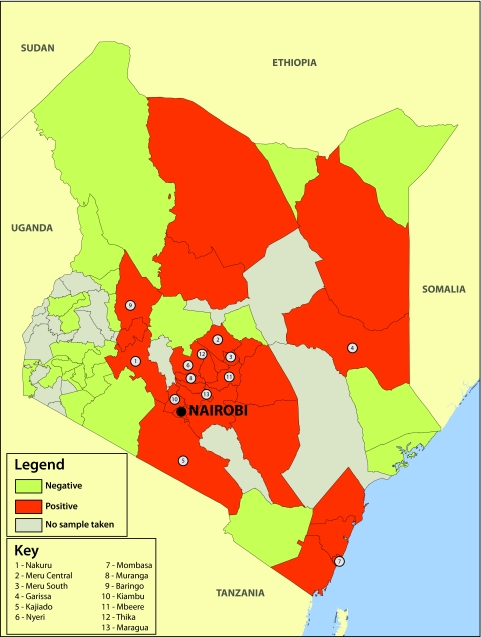
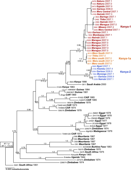
Rift Valley fever (RVF) virus historically has caused widespread and extensive outbreaks of severe human and livestock disease throughout Africa, Madagascar, and the Arabian Peninsula. Following unusually heavy rainfall during the late autumn of 2006, reports of human and animal illness consistent with RVF virus infection emerged across semiarid regions of the Garissa District of northeastern Kenya and southern Somalia. Following initial RVF virus laboratory confirmation, a high-throughput RVF diagnostic facility was established at the Kenyan Central Veterinary Laboratories in Kabete, Kenya, to support the real-time identification of infected livestock and to facilitate outbreak response and control activities. A total of 3,250 specimens from a variety of animal species, including domesticated livestock (cattle, sheep, goats, and camels) and wildlife collected from a total of 55 of 71 Kenyan administrative districts, were tested by molecular and serologic assays. Evidence of RVF infection was found in 9.2% of animals tested and across 23 districts of Kenya, reflecting the large number of affected livestock and the geographic extent of the outbreak. The complete S, M, and/or L genome segment sequence was obtained from a total of 31 RVF virus specimens spanning the entire known outbreak period (December-May) and geographic areas affected by RVF virus activity. Extensive genomic analyses demonstrated the concurrent circulation of multiple virus lineages, gene segment reassortment, and the common ancestry of the 2006/2007 outbreak viruses with those from the 1997-1998 east African RVF outbreak. Evidence of recent increases in genomic diversity and effective population size 2 to 4 years prior to the 2006-2007 outbreak also was found, indicating ongoing RVF virus activity and evolution during the interepizootic/epidemic period. These findings have implications for further studies of basic RVF virus ecology and the design of future surveillance/diagnostic activities, and they highlight the critical need for safe and effective vaccines and antiviral compounds to combat this significant veterinary and public health threat.


Figures

FIG. 1.
FIG. 1.
Administrative district-level map of the Republic of Kenya. Shaded in red are districts in which evidence of acute-phase infection (by qRT-PCR, RVF virus antigen capture ELISA, and/or anti-RVF virus-specific IgM ELISA) was found.
FIG. 2.
FIG. 2.
RVF virus diagnostic assay performance. Results of serologic testing are reported as the adjusted SUMOD, which was defined as the cumulative sum of the optical densities of each specimen dilution minus the background absorbance of uninfected control Vero E6 cells. (A) Pairwise comparison of the qRT-PCR CT values (y axis) and RVF antigen capture ELISA adjusted SUMOD values (x axis). qRT-PCR CT values of less than 40 were considered positive. The cutoff SUMOD value for the RVF antigen capture ELISA was conservatively set at >0.45 and is indicated by the dashed red line and arrow. (B) Pairwise comparison of the qRT-PCR CT values (y axis) and anti-RVF virus IgM ELISA adjusted SUMOD values (x axis). qRT-PCR CT values of less than 40 were considered positive. The cutoff SUMOD value for the anti-RVF virus specific IgM ELISA was conservatively set at >0.75 and is indicated by the dashed red line and arrow. Note the marked reduction in qRT-PCR-positive (n = 3) specimens after individual animals became seropositive for anti-RVF virus-specific IgM. (C) Pairwise comparison of the qRT-PCR CT values (y axis) and anti-RVF virus IgG ELISA adjusted SUMOD values (x axis). qRT-PCR CT values of less than 40 were considered positive. The cutoff SUMOD value for the anti-RVF virus specific IgG ELISA was conservatively set at >1.50 and is indicated by the dashed red line and arrow. (D) Pairwise comparison of anti-RVF virus IgM ELISA adjusted SUMOD values (y axis) and anti-RVF virus IgG ELISA adjusted SUMOD values (x axis) of all RVF virus-positive specimens (recent and past). The cutoff values of the IgM and IgG ELISAs are indicated by the red horizontal dashed line and green vertical dashed line, respectively. The resulting chart is divided into quadrants, with the upper left quadrant indicating specimens that were positive only for IgM serology, the upper right quadrant indicating specimens that were both IgM and IgG positive, and the lower left quadrant indicating specimens positive by qRT-PCR only.
FIG. 3.
FIG. 3.
RVF virus S segment maximum a posteriori clade credibility tree generated using BEAST-v1.4.7/Tree annotator/Fig Tree-v1.1.2. The combined MCMC chain length was 9.0 × 10⁷ steps; 2.25 × 10⁷ steps (25%) were removed as the burn in. Posterior support values (HPD) are indicated as integers (i.e., 100% support = 1.0) above each node. The calculated mean TMRCAs are indicated below each respective node and are enumerated as years before the collection date of the last outbreak specimen (May 2007). Note that 31 individual complete RVF virus S segments were obtained. The main Kenya-1 lineage is indicated in red, with the sublineage Kenya-1a in orange and the separate Kenya-2 lineage depicted in blue.
FIG. 4.
FIG. 4.
RVF virus M segment maximum a posteriori clade credibility tree generated using BEAST-v1.4.7/Tree annotator/Fig Tree-v1.1.2. The combined MCMC chain length was 9.0 × 10⁷ steps; 2.25 × 10⁷ steps (25%) were removed as the burn in. Posterior support values (HPD) are indicated as integers (i.e., 100% support = 1.0) above each node. The calculated mean TMRCAs are indicated below each respective node and are enumerated as years before the collection date of the last outbreak specimen (May 2007). Note that 27 individual complete RVF virus M segments were obtained. The main Kenya-1 lineage is indicated in red, with the sublineage Kenya-1a in orange and the separate Kenya-2 lineage depicted in blue.
FIG. 5.
FIG. 5.
RVF virus L segment maximum a posteriori clade credibility tree generated using BEAST-v1.4.7/Tree annotator/Fig Tree-v1.1.2. The combined MCMC chain length was 9.0 × 10⁷ steps; 2.25 × 10⁷ steps (25%) were removed as the burn in. Posterior support values (HPD) are indicated as integers (i.e., 100% support = 1.0) above each node. The calculated mean TMRCAs are indicated below each respective node and are enumerated as years before the collection date of the last outbreak specimen (May 2007). Note that 27 individual complete RVF virus L segments were obtained. The main Kenya-1 lineage is indicated in red, with the sublineage Kenya-1a in orange and the separate Kenya-2 lineage depicted in blue.
FIG. 6.
FIG. 6.
MSNs visually describing discrete genetic distances between unique haplotypes for the S, M, and L RVF virus genome segments. Each node represents one nucleotide difference between extant (open circle) or inferred (filled black circle) haplotypes. Proportionally larger open circles or squares represent the relative number of extant haplotypes represented in the network. Note the greater distance measured in nucleotide changes (steps) between the Kenya-1 and Kenya-2 lineages than that with the prototype Kenyan 1997-1998 RVF virus strain. Also note the star-like phylogeny of the Kenya-1 lineage, indicating the potential of recent increases in virus population size or geographic range. An asterisk on each MSN indicates the relative position of the putative M segment reassortant virus (#0608).
FIG. 7.
FIG. 7.
Mismatch distributions of the Kenya-1 and Kenya-2 lineage S, M, and L RVF virus genome segments. In each panel, the mismatch distributions of the Kenya-1 lineage are depicted on the left, with the Kenya-2 mismatch distributions depicted on the right. The actual observed frequencies of nucleotide (nt) substitutions between unique haplotypes are depicted in blue with diamond data markers. Simulated data of recent exponential population demographic expansion are depicted in red with square data markers. Analyses of the S segment were clearly unimodal for lineage Kenya-1, allowing for the rejection of the null hypothesis of stable population size and spatial expansion (P = 0.57 and 0.61, respectively) and providing evidence that an increase in virus population size and spatial distribution had occurred. Mismatch distributions for lineage Kenya-1 based on the M and L segments appeared to be multimodal, but no significant differences were found between the observed data and that simulated under a model of sudden expansion (M, P = 0.94 and 0.89; L, P = 0.70 and 0.62). Based on these results, the evidence of recent exponential population expansion prior to the 2006-2007 outbreak could not be ruled out for the S, M, and L RVF virus genome segments of the Kenya-1 lineage.

Similar articles

Cited by

References

    1. Anonymous. 2007. Outbreaks of Rift Valley fever in Kenya, Somalia and United Republic of Tanzania, December 2006-April 2007. Wkly. Epidemiol. Rec. 82169-178. - PubMed
    1. Anyamba, A., J. P. Chretien, J. Small, C. J. Tucker, and K. J. Linthicum. 2006. Developing global climate anomalies suggest potential disease risks for 2006-2007. Int. J. Health Geogr. 560. - PMC - PubMed
    1. Beaty, B. J., E. J. Rozhon, P. Gensemer, and D. H. Bishop. 1981. Formation of reassortant bunyaviruses in dually infected mosquitoes. Virology 111662-665. - PubMed
    1. Bird, B. H., C. G. Albarino, and S. T. Nichol. 2007. Rift Valley fever virus lacking NSm proteins retains high virulence in vivo and may provide a model of human delayed onset neurologic disease. Virology 36210-15. - PubMed
    1. Bird, B. H., D. A. Bawiec, T. G. Ksiazek, T. R. Shoemaker, and S. T. Nichol. 2007. Highly sensitive and broadly reactive quantitative reverse transcription-PCR assay for high-throughput detection of Rift Valley fever virus. J. Clin. Microbiol. 453506-3513. - PMC - PubMed

Publication types

MeSH terms

LinkOut - more resources