Comparative Study
. 2008 Nov;82(22):11344-53.
doi: 10.1128/JVI.02375-07. Epub 2008 Sep 10.

Structural evolution of reoviridae revealed by oryzavirus in acquiring the second capsid shell

Affiliations
Comparative Study

Structural evolution of reoviridae revealed by oryzavirus in acquiring the second capsid shell

Naoyuki Miyazaki et al. J Virol. 2008 Nov.

Abstract

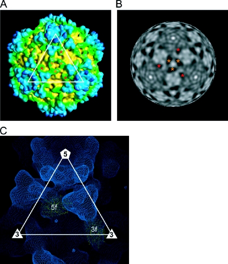
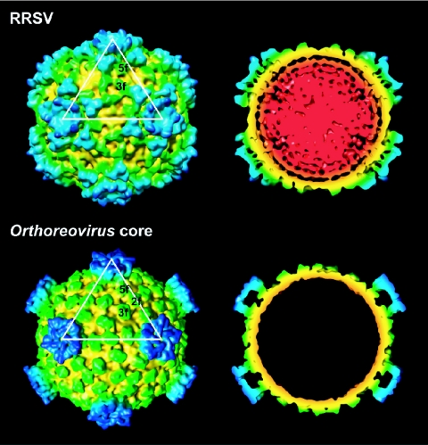
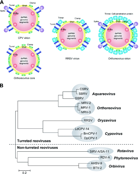
The conservation of the core structure and diversification of the external features among the turreted reoviruses appear to be relevant to structural evolution in facilitating the infection of diverse host species. The structure of Rice ragged stunt virus (RRSV), in the genus Oryzavirus of the family Reoviridae, is determined to show a core composed of capsid shell, clamps, and long turrets. The RRSV core structure is equivalent to the core structure of Orthoreovirus and the virion structure of Cytoplasmic polyhedrosis virus (CPV). In RRSV, five peripheral trimers surround each long turret and sit at the Q trimer position in the T=13l icosahedral symmetry, a structural feature unique to turreted reoviruses. That is, the core of RRSV is partially covered by 60 copies of the peripheral trimer. In contrast, the core of Orthoreovirus is covered by 200 copies of the trimer that sit at the Q, R, S, and T trimer positions. Our results suggest that among the three viruses, RRSV has a structure intermediate between that of Orthoreovirus and the CPV virion. This conclusion coincides with the results of the phylogenetic analysis of amino acid sequences of RNA-dependent RNA polymerases.


Figures

FIG. 1.
FIG. 1.
(A) Electron micrograph of uranyl acetate-stained RRSV (upper) and projection images calculated from the 3D reconstruction (lower). The bar represents 100 nm. (B) Sodium dodecyl sulfate-10% polyacrylamide gel electrophoresis of proteins from particles of RRSV (lane 1) and RDV (lane 2). Positions of RDV protein species are shown. (C) Electron micrograph of RRSV embedded in vitreous ice. The image was recorded at an applied underfocus value of 3.0 μm with a cryoelectron micrograph (JEM-2100F) operated at 200 kV and a nominal magnification of ×30,000. Empty particles are indicated by the arrows. The bar represents 100 nm. (D) Resolution assessment. The Fourier shell correlation coefficient is plotted.
FIG. 2.
FIG. 2.
Overall structure of RRSV. (A) Surface representation of RRSV. (B) Central cross-section (40 Å thick). The map is colored according to the distance from the center of the viral particle, for which color coding is indicated. Local structures are visible as follows: core capsid, yellow; clamp proteins, green; long turret, blue structures located at the fivefold axes; and peripheral trimers, blue trimers located around turrets.
FIG. 3.
FIG. 3.
Turret structure. (A) Close-up view of a turret. The two globular domains that are marked by asterisks might correspond to two methylase domains. (B) Radially cued density viewed along an icosahedral fivefold axis at a radius of 343 Å showing the molecular boundaries of the five subunits in a turret. High to low densities are indicated by a gray scale. (C) Atomic structure of the Orthoreovirus λ2 turret. A CPK model of a pentameric turret is shown in the upper panel. Five subunits are shown in different colors. Asterisks indicate methylase domains as described for panel A. The lower panel shows ribbon drawings of the monomer structure of the turret protein. The GTase, methylase-1, methylase-2, and immunoglobulin-like domains are colored in red, yellow, green, and cyan, respectively.
FIG. 4.
FIG. 4.
Clamp proteins. (A and B) Surface representation (A) and radially cued density at a radius of 293 Å (B), viewed along a threefold axis. Two icosahedrally independent 3f and 5f clamp proteins are marked with orange and red asterisks, respectively. (C) The atomic structures of the clamp σ2 protein of Orthoreovirus were fitted into the cryo-EM map of RRSV.
FIG. 5.
FIG. 5.
Structural comparison between the RRSV virion and the Orthoreovirus core. Surface representations and central cross-sections (40 Å thick) of the RRSV virion and the Orthoreovirus core are shown. The color coding is the same as that described in the legend to Fig. 1.
FIG. 6.
FIG. 6.
Peripheral trimers. (A) Close-up view of a trimer. A peripheral trimer is shown bound to three clamps that are marked with asterisks. (B) Radially cued density with fivefold axes at a radius of 333 Å, at which the molecular boundaries of trimers are apparent. (C) Atomic structures of the Orthoreovirus trimers. CPK models of σ33 (left) and μ13σ33 trimers (right) are shown as viewed from the outside of the capsid shell (upper) and after 90° rotation (lower). The σ3 molecules are colored pink, magenta, and orange, and the μ1 molecules are colored cyan, blue, and green. The σ3 molecules are removed from the viral particles during the infection by proteolysis. (D) Surface representation with the icosahedral T=13l net. The positions of trimers that correspond to the Q position in the icosahedral T=13l symmetry are shown and labeled Q.
FIG. 7.
FIG. 7.
Relationships among turreted reoviruses. (A) Schematic representation of the RRSV virion, the CPV virion, the Orthoreovirus core, and the Orthoreovirus virion. The RdRp, capsid, clamp, trimeric outer capsid, turret, and spike proteins are shown in red, yellow, green, sky blue, blue, and blue-purple, respectively. (B) Neighbor-joining tree constructed from full-length sequences of RdRps of viruses in the family Reoviridae. The genera, strains, abbreviations of names of viruses, and accession numbers of sequences are as follows. For Aquareovirus, Chum salmon reovirus (CSRV; AF418295), Striped bass reovirus (SBRV; AF450318), and Golden shiner reovirus (GSRV; AF403399); for Orthoreovirus, Mammalian orthoreovirus subgroup 1, Lang strain (MRV-1; M24734), Jones strain (MRV-2; M31057), and Dearing strain (MRV-3; M31058); for Oryzavirus, Rice ragged stunt virus (RRSV; U66714); for Cypovirus, Bombyx mori cytoplasmic polyhedrosis virus 1 (BmCPV-1; AF323782), Dendrolimus punctatus cypovirus 1 (DpCPV-1; AAN46860), and Lymantria dispar cypovirus 14 (LdCPV-14; AAK73087); for Rotavirus, Rotavirus A and Simian rotavirus strain SA11 (SiRV-A/SA-11; AF015955); for Phytoreovirus, Rice dwarf virus strain A (RDV-A; D90198); and for Orbivirus, African horse sickness virus serotype 9 (AHSV-9; U94887) and Bluetongue virus serotype 2 (BTV-2; L20508) (1).

Similar articles

Cited by

References

    1. Attoui, H., F. Mohd Jaafar, M. Belhouchet, P. Biagini, J. F. Cantaloube, P. de Micco, and X. de Lamballerie. 2005. Expansion of family Reoviridae to include nine-segmented dsRNA viruses: isolation and characterization of a new virus designated Aedes pseudoscutellaris reovirus assigned to a proposed genus (Dinovernavirus). Virology 343212-223. - PubMed
    1. Baker, T. S., and R. H. Cheng. 1996. A model-based approach for determining orientations of biological macromolecules imaged by cryoelectron microscopy. J. Struct. Biol. 116120-130. - PubMed
    1. Bamford, D. H., J. M. Grimes, and D. I. Stuart. 2005. What does structure tell us about virus evolution? Curr. Opin. Struct. Biol. 15655-663. - PubMed
    1. Bartlett, N., S. Gillies, S. Bullivant, and A. Bellamy. 1974. Electron microscopy study of reovirus reaction cores. J. Virol. 14315-326. - PMC - PubMed
    1. Bellamy, A., and J. Harvey. 1976. Biophysical studies of reovirus type 3. III. A laser light-scattering study of the RNA transcriptase reaction. Virology 7028-36. - PubMed

Publication types

LinkOut - more resources