. 2009 Jan;150(1):187-99.
doi: 10.1210/en.2008-0368. Epub 2008 Sep 11.

Role of dosage-sensitive sex reversal, adrenal hypoplasia congenita, critical region on the X chromosome, gene 1 in protein kinase A- and protein kinase C-mediated regulation of the steroidogenic acute regulatory protein expression in mouse Leydig tumor cells: mechanism of action

Affiliations

Role of dosage-sensitive sex reversal, adrenal hypoplasia congenita, critical region on the X chromosome, gene 1 in protein kinase A- and protein kinase C-mediated regulation of the steroidogenic acute regulatory protein expression in mouse Leydig tumor cells: mechanism of action

Pulak R Manna et al. Endocrinology. 2009 Jan.

Abstract

Dosage-sensitive sex reversal, adrenal hypoplasia congenita, critical region on the X chromosome, gene 1 (DAX-1) is an orphan nuclear receptor that has been demonstrated to be instrumental to the expression of the steroidogenic acute regulatory (StAR) protein that regulates steroid biosynthesis in steroidogenic cells. However, its mechanism of action remains obscure. The present investigation was aimed at exploring the molecular involvement of DAX-1 in protein kinase A (PKA)- and protein kinase C (PKC)-mediated regulation of StAR expression and its concomitant impact on steroid synthesis using MA-10 mouse Leydig tumor cells. We demonstrate that activation of the PKA and PKC pathways, by a cAMP analog dibutyryl (Bu)2cAMP [(Bu)2cAMP] and phorbol 12-myristate 13-acetate (PMA), respectively, markedly decreased DAX-1 expression, an event that was inversely correlated with StAR protein, StAR mRNA, and progesterone levels. Notably, the suppression of DAX-1 requires de novo transcription and translation, suggesting that the effect of DAX-1 in regulating StAR expression is dynamic. Chromatin immunoprecipitation studies revealed the association of DAX-1 with the proximal but not the distal region of the StAR promoter, and both (Bu)2cAMP and PMA decreased in vivo DAX-1-DNA interactions. EMSA and reporter gene analyses demonstrated the functional integrity of this interaction by showing that DAX-1 binds to a DNA hairpin at position -44/-20 bp of the mouse StAR promoter and that the binding of DAX-1 to this region decreases progesterone synthesis by impairing transcription of the StAR gene. In support of this, targeted silencing of endogenous DAX-1 elevated basal, (Bu)2cAMP-, and PMA-stimulated StAR expression and progesterone synthesis. Transrepression of the StAR gene by DAX-1 was tightly associated with expression of the nuclear receptors Nur77 and steroidogenic factor-1, demonstrating these factors negatively modulate the steroidogenic response. These findings provide insight into the molecular events by which DAX-1 influences the PKA and PKC signaling pathways involved in the regulation of the StAR protein and steroidogenesis in mouse Leydig tumor cells.


Figures
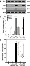
Figure 1
Figure 1
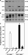
Role of PKA and PKC signaling in DAX-1, StAR, P-StAR, and progesterone levels in MA-10 cells. Cells were treated with two different concentrations of (Bu)2cAMP (0.1 and 0.5 mm) and PMA (1 and 10 nm), respectively, and subjected to preparation of whole cell lysates for Western blot analysis using 20–30 μg of protein. Representative immunoblots illustrate expression of DAX-1, StAR, and P-StAR in different treatment groups (A). Integrated OD (IOD) values of each band were quantified and compiled data (n = 4) are presented (B). C, Accumulation of progesterone in the media of the same treatment groups was determined and expressed as nanograms per milligram protein. Inset, Control (Con) and PMA (10 nm)-treated progesterone levels. Data represent the mean ± se of four independent experiments. Letters above the bars indicate that these groups differ significantly from each other, at least at P < 0.05.
Figure 2
Figure 2
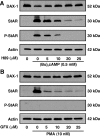
Inhibition of PKA and PKC signaling on DAX-1, StAR, and P-StAR levels in MA-10 cells. Cells were pretreated with increasing doses PKA (H89; 0–25 μm) and PKC (GFX; 0–25 μm) inhibitors for 30 min and then incubated without or with (Bu)2cAMP (0.5 mm; A) and PMA (10 nm; B) for an additional 6 h. Actin expression was assessed as a loading control (A and B). Representative immunoblots illustrate expression of DAX-1, StAR, and P-StAR using 25–30 μg of total protein. Data are representative of three independent experiments.
Figure 3
Figure 3
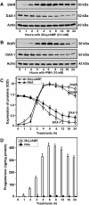
Time-course expression patterns of StAR and DAX-1 proteins and progesterone synthesis in response to (Bu)2cAMP (0.5 mm) and PMA (10 nm). MA-10 cells were treated for 0–24 h at the indicated doses of (Bu)2cAMP (A) and PMA (B), and 22–25 μg of total protein were used for Western blot analyses. Representative immunoblots illustrate the expression of StAR and DAX-1 in different treatment groups. Integrated OD (IOD) values for each band were quantified and compiled data from three independent experiments for both proteins are presented (C). Actin expression was assessed as a loading control. Accumulation of progesterone in the media of the same treatment groups was determined and expressed as nanograms per milligram protein, which represent the mean ± se of three independent experiments (D). Asterisks indicate the first time point at which values were significantly different (P < 0.05) from controls.
Figure 4
Figure 4
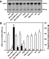
Inhibition of DAX-1 expression by PKA and PKC signaling requires transcription and de novo protein synthesis. MA-10 cells were treated without (Con) or with (Bu)2cAMP (0.5 mm) and PMA (10 nm) in the absence or presence of Act (5 μm) and Chx (5 μm) for 6 h and subjected to DAX-1 mRNA expression by RT-PCR analysis (A). A representative autoradiogram illustrates the expression of DAX-1 and L19 in different treatment groups using 2 μg of total RNA. Integrated OD (IOD) values of each band were quantified and normalized to the corresponding L19 bands (B). Accumulation of progesterone in the media was determined and expressed as nanograms per milligram protein (B). Results represent the mean ± se of three independent experiments. Note the different scales on the graph. Letters above the bars indicate that these groups differ significantly from each other at least at P < 0.05. *, P < 0.05; **, P < 0.01; ***, P < 0.001 vs. control DAX-1 mRNA.
Figure 5
Figure 5
Effects of PKA and PKC on the association of DAX-1 with the StAR promoter. MA-10 cells were treated without or with increasing doses of (Bu)2cAMP (0–1.0 mm, A) and PMA (0–20 nm, B) for 6 h, and ChIP assays were carried out as described in Materials and Methods. Cross-linked sheared chromatin obtained from (Bu)2cAMP and PMA-treated cells was immunoprecipitated (IP) with either IgG or anti-DAX-1 and anti-P-CREB (D) Abs. Recovered chromatin was subjected to PCR analysis using primers (−170/−1 bp, A, B, and D) encompassing the DAX-1 binding site or primers (3596/−3428 bp, C) approximately 3500 bp upstream of the DAX-1 region. Representative autoradiograms illustrate the association of DAX-1 and P-CREB with the StAR promoter in response to (Bu)2cAMP and PMA. Data shown are representative of three independent experiments.
Figure 6
Figure 6
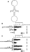
Identification of a DNA hairpin structure in the mouse StAR promoter and the effect of DAX-1 on PKA- and PKC-mediated StAR promoter activity. A, Presence of a putative DAX-1 binding motif in the mouse StAR gene. This motif is located between −44 and −20 bp and is composed of five nucleotides stem and 15 nucleotides loop. B, MA-10 cells were transfected with different StAR reporter plasmids (−151/−1 and −68/−1 bp) in the presence of 20 ng of pRL-SV40 vector (renilla luciferase for determining transfection efficiency). Schematic representations of different StAR reporters (−151 and −68 bp) are shown (bottom panel). pGL3 basic (pGL3) was used as a control. Cells were also transfected with empty vector (pcDNA3) or DAX-1 expression plasmid, within the context of the −151/−1 wild-type and mutant StAR reporter segments as indicated, in the presence of pRL-SV40 (C). After 36 h of transfection, cells were incubated for a further 6 h in the absence (basal) or presence of either (Bu)2cAMP (0.5 mm) or PMA (10 nm), and luciferase activity in the cell lysates was determined and expressed as relative light units (RLU, luciferase/renilla). Relative fold activity is illustrated above bar diagrams. Data represent the mean ± se of four independent experiments. Letters above the bars indicate that these groups differ significantly from each other at least at P < 0.05.
Figure 7
Figure 7
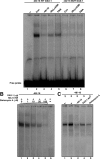
The binding of the hairpin region in the StAR promoter to endogenous DAX-1 in MA-10 NE and in vitro-translated DAX-1 using EMSAs. MA-10 NE (12–15 μg) obtained from control (Con, lanes 2, 3, and 6), 0.5 mm (Bu)2cAMP (lanes 4 and 7), and 10 nm PMA (lanes 5 and 8)-treated cells were incubated with the 32P-labeled wild-type (WT) and Mut DAX-1 probes (−48/−16 bp) encompassing the hairpin loop (A) as described in Materials and Methods. The specificity of DAX-1 binding (−48/−16 bp) in untreated MA-10 NE (lanes 1–6) was also assessed with increasing amounts (1–100 μm) of distamycin A (B). Binding of in vitro-translated DAX-1 protein (lanes 1–5) was evaluated with the −48/−16 probe (C). Protein-DNA binding was augmented with an increasing amount of DAX-1 (lanes 1–5). Binding was challenged with cold competitor (lane 3), DAX-1 Ab (lane 4), and distamycin A (lane 5). Cold competitor (−48/−16 bp) was used at 100-fold molar excess. Migration of free probes is shown in A. These experiments were repeated three times, and representative phosphor images from each group are illustrated.
Figure 8
Figure 8
Silencing of DAX-1 and its relevance to StAR expression and progesterone synthesis. MA-10 cells were transfected with either a negative control siRNA at 100 nm (Con siRNA) or a mixture of two DAX-1-specific siRNAs at 50 nm (100 nm total, DAX-1 siRNA), as described in Materials and Methods. After 36 h of transfection, cells were treated without (Con) or with (Bu)2cAMP (0.5 mm) and PMA (10 nm) for an additional 6 h and subjected to Western (DAX-1, StAR, and actin) and RT-PCR (StAR, DAX-1, and L19) analyses. Representative immunoblots and autoradiograms illustrate the expression of DAX-1, StAR, and actin (A) and StAR, DAX-1, and L19 (B) in different treatment groups, respectively. Actin and L19 were used as controls in Western and RT-PCR analyses, respectively. Similar results were obtained from three different experiments. C, Accumulation of progesterone in the media was determined and expressed as nanograms per milligram protein, and the data shown represent the mean ± se of three independent experiments. Letters above the bars indicate that these groups differ significantly from each other, at least at P < 0.05.
Figure 9
Figure 9
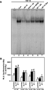
Assessment of Nur77 and SF-1 binding to the DAX-1 hairpin structure and roles of these factors in StAR promoter responsiveness. A, MA-10 NE (15 μg) was used to determine protein binding to the 32P-labeled DAX-1 (−48/−16 bp) motif (lanes 2–7). Binding of untreated MA-10 NE (Con, lane 2) to the labeled probe (−48/−16) was challenged with its unlabeled (−48/−16 bp) sequence (lane 3), DAX-1 Ab (lane 4), consensus NBRE (Con NBRE, lane 5), a confirmed SF-1 BS (lane 6), and with a mutant DAX-1 sequence (−48/−16 Mut, lane 7). MA-10 cells were transfected with empty vector (pcDNA3), Nur77, and SF-1 expression plasmids, within the context of one of the −151/−1 StAR reporter segments: either wild-type (−151 WT) or a reporter containing mutations in one of the SF-1 binding sites (SF-1/1M, SF-1/2M, SF-1/3M), in the presence of 15–20 ng of pRL-SV40 (B). After 48 h of transfection, cells were collected and luciferase activity in the cell lysates determined by [relative light units (RLU), luciferase/renilla] and presented in terms of percent activity. Data represent the mean ± se of four independent experiments. Letters above the bars indicate that these groups differ significantly from each other, at least at P < 0.05.
Figure 10
Figure 10
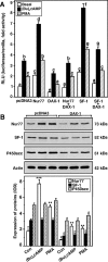
The effects of the expression of Nur77, SF-1, and DAX-1, independently or in combination on StAR promoter activity; the effects of DAX-1, (Bu)2cAMP, and PMA on Nur77, SF-1; and P450scc expression in MA-10 cells. A, Using the −151/−1-bp StAR reporter segment, cells were transfected with empty vector (pcDNA3), Nur77, SF-1, DAX-1, or a combination thereof as indicated in the presence of 15–20 ng of pRL-SV40. After 36 h of transfection, cells were incubated for an additional 6 h in the absence (basal) or presence of (Bu)2cAMP (0.5 mm) and PMA (10 nm), and luciferase activity in the cell lysates was determined [relative light units (RLU), luciferase/renilla] and expressed in terms of fold activity. Data represent the mean ± se of three to five independent experiments. Cells were also transfected with either empty vector (pcDNA3) or DAX-1 expression plasmids and subjected to immunoblotting for Nur77, SF-1, and P450scc expression using 20–25 μg of total cellular protein (B). Cells were treated with (Bu)2cAMP (0.5 mm) and PMA (10 nm) for 6 h, and the expression of Nur77, SF-1, and P450scc proteins was determined by immunoblotting. Actin expression demonstrates equal loading. Representative immunoblots illustrate the expression of Nur77, SF-1, and P450scc proteins in different treatment groups. Integrated OD (IOD) values of each band were quantified and compiled data from three independent experiments are presented (lower panel). Letters above the bars indicate that these groups differ significantly from each other, at least at P < 0.05. *, P < 0.05; **, P < 0.01; ***, P < 0.001 represent significant differences in comparison with respective controls.

Similar articles

Cited by

References

    1. Zanaria E, Muscatelli F, Bardoni B, Strom TM, Guioli S, Guo W, Lalli E, Moser C, Walker AP, McCabe ER, Meitinger T, Monaco AP, Sassone-Corsi P, Camerino G An unusual member of the nuclear hormone receptor superfamily responsible for X-linked adrenal hypoplasia congenita. Nature 372:635–641 - PubMed
    1. Muscatelli F, Strom TM, Walker AP, Zanaria E, Recan D, Meindl A, Bardoni B, Guioli S, Zehetner G, Rabl W, Schwarz HP, Kaplan J-C, Camerino G, Meitinger T, Monaco AP 1994 Mutations in the DAX-1 gene give rise to both X-linked adrenal hypoplasia congenita and hypogonadotropic hypogonadism. Nature 372:672–676 - PubMed
    1. Tamai KT, Monaco L, Alastalo TP, Lalli E, Parvinen M, Sassone-Corsi P 1996 Hormonal and developmental regulation of DAX-1 expression in Sertoli cells. Mol Endocrinol 10:1561–1569 - PubMed
    1. Reutens AT, Achermann JC, Ito M, Gu WX, Habiby RL, Donohoue PA, Pang S, Hindmarsh PC, Jameson JL 1999 Clinical and functional effects of mutations in the DAX-1 gene in patients with adrenal hypoplasia congenita. J Clin Endocrinol Metab 84:504–511 - PubMed
    1. Meeks JJ, Weiss J, Jameson JL 2003 Dax1 is required for testis determination. Nat Genet 34:32–33 - PubMed

Publication types

MeSH terms