Review
. 2008 Nov 1;586(21):5047-61.
doi: 10.1113/jphysiol.2008.160440. Epub 2008 Sep 11.

Smooth muscle cell calcium activation mechanisms

Affiliations
Review

Smooth muscle cell calcium activation mechanisms

Michael J Berridge. J Physiol. .

Abstract

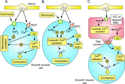
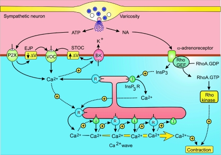
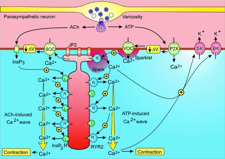
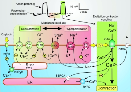
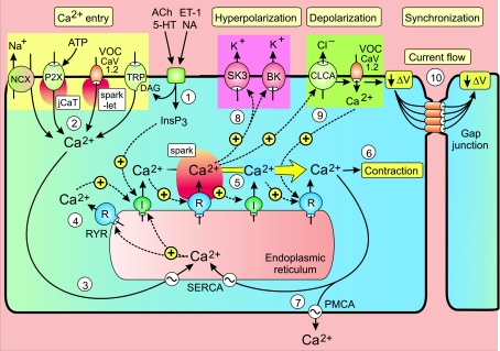
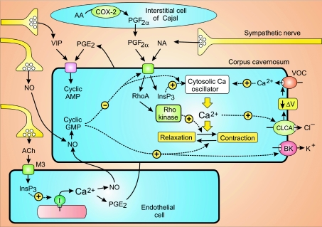
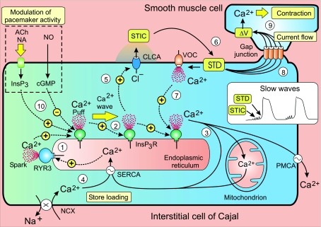
Smooth muscle cell (SMC) contraction is controlled by the Ca2+ and Rho kinase signalling pathways. While the SMC Rho kinase system seems to be reasonably constant, there is enormous variation with regard to the mechanisms responsible for generating Ca2+ signals. One way of dealing with this diversity is to consider how this system has been adapted to control different SMC functions. Phasic SMCs (vas deferens, uterus and bladder) rely on membrane depolarization to drive Ca2+ influx across the plasma membrane. This depolarization can be induced by neurotransmitters or through the operation of a membrane oscillator. Many tonic SMCs (vascular, airway and corpus cavernosum) are driven by a cytosolic Ca2+ oscillator that generates periodic pulses of Ca2+. A similar oscillator is present in pacemaker cells such as the interstitial cells of Cajal (ICCs) and atypical SMCs that control other tonic SMCs (gastrointestinal, urethra, ureter). The changes in membrane potential induced by these cytosolic oscillators does not drive contraction directly but it functions to couple together individual oscillators to provide the synchronization that is a characteristic feature of many tonic SMCs.


Figures

Figure 1
Figure 1. The three main mechanisms responsible for generating the Ca2+ transients that trigger smooth muscle cell (SMC) contraction
A, receptor-operated channels (ROCs) or a membrane oscillator induces the membrane depolarization (ΔV) that triggers Ca2+ entry and contraction. B, a cytosolic Ca2+ oscillator induces the Ca2+ signal that drives contraction. C, a cytosolic Ca2+ oscillator in interstitial cells of Cajal (ICCs) or atypical SMCs induces the membrane depolarization that spreads through the gap junctions to activate neighbouring SMCs. Reproduced from Berridge (2008), with permission.
Figure 2
Figure 2. Neural activation of vas deferens smooth muscle cells
Sympathetic neurons have numerous varicosities that release ATP and noradrenaline (NA) that activate contraction through separate signalling pathways. See text for further details. BK, large conductance Ca2+-sensitive K+ channels; EJP, excitatory junction potential; STOC, spontaneous transient outward currents; I, inositol 1,4,5-trisphosphate (InsP3) receptor; R, ryanodine receptor. Reproduced from Berridge (2008), with permission.
Figure 3
Figure 3. Bladder smooth muscle cells (SMCs) have a membrane oscillator that generates the periodic action potentials that initiate the process of excitation–contraction coupling
Neurotransmitters such as ATP and acetylcholine (ACh), which are released from parasympathetic axonal varicosities that innervate the bladder, activate or accelerate the oscillator by inducing membrane depolarization (ΔV) through two separate pathways. Reproduced from Berridge (2008), with permission.
Figure 4
Figure 4. A model for the activation mechanism of human uterus contractility
Contractions of the uterus during labour are driven by an endogenous membrane oscillator (horizontal green to pink box) that induces the slow pacemaker depolarization responsible for triggering the excitation–contraction coupling mechanism (vertical green box). The oscillator depends on an interaction between ion channels, pumps and exchangers that are colour coordinated to indicate their contribution to either depolarization (green) or hyperpolarization (red). When the pacemaker depolarization (ΔV) reaches the threshold for the activation of the L-type voltage-operated channels (VOCs) (dashed line in the membrane potential trace at the top), excitation–contraction coupling is triggered and the muscle contracts. See text for further details. Reproduced from Berridge (2008), with permission.
Figure 5
Figure 5. Vascular or airway SMCs are driven by a cytosolic oscillator that generates a periodic release of Ca2+ from the endoplasmic reticulum that usually appears as a propagating Ca2+ wave
The oscillator is induced/modulated by neurotransmitters such as acetylcholine (ACh), 5-hydroxytryptamine (5-HT), noradrenaline (NA) and endothelin-1 (ET-1), which act through inositol 1,4,5-trisphosphate (InsP3) that initiates the oscillatory mechanism. The sequence of steps 1–9 is described in the text. Reproduced from Berridge (2008), with permission.
Figure 6
Figure 6. The corpus cavernosum SMCs, which are poised between contraction and relaxation, are controlled by multiple stimuli released from neurons, endothelial cells and the interstitial cells of Cajal
Some of the stimuli such as noradrenaline (NA) and prostaglandin F (PGF) drive contraction, whereas nitric oxide (NO), prostaglandin E2 (PGE2) and vasoactive intestinal peptide (VIP) control relaxation. See text for details of their signalling pathways. Reproduced from Berridge (2008), with permission.
Figure 7
Figure 7. The cytosolic Ca2+ oscillator responsible for pacemaker activity in interstitial cells of Cajal releases periodic pulses of Ca2+ that form a Ca2+ wave
The increase in Ca2+ activates Cl channels (CLCA) to give the spontaneous transient inward currents (STICs) that sum to form the spontaneous transient depolarizations (STD) resulting in the slow waves of membrane depolarization (see inset). Current flow through gap junctions allows these waves to spread into neighbouring smooth muscle cells to activate contraction. See text for a description of the oscillator that drives this activation process. Reproduced from Berridge (2008), with permission.

References

    1. Amberg GC, Navedo MF, Nieves-Cintrón M, Molkentin JD, Santana LF. Calcium sparklets regulate local and global calcium in murine arterial smooth muscle. J Physiol. 2007;579:187–201. - PMC - PubMed
    1. Aoyama M, Yamada A, Wang J, Ohya S, Furuzono S, Goto T, Hotta S, Ito Y, Matsubara T, Shimokata K, Chen SRW, Imaizumi Y, Nakayama S. Requirement of ryanodine receptors for pacemaker Ca2+ activity in ICC and HEK293 cells. J Cell Sci. 2004;117:2813–2825. - PubMed
    1. Berridge MJ. Inositol trisphosphate and calcium oscillations. Biochem Soc Symp. 2007;74:1–7. - PubMed
    1. Berridge MJ. Cell Signalling Biology. Portland Press Limited; 2008. http://www.cellsignallingbiology.org.
    1. Berridge MJ, Bootman MD, Roderick HL. Calcium signalling: Dynamics, homeostasis and remodelling. Nat Rev Mol Cell Biol. 2003;4:517–529. - PubMed

LinkOut - more resources