. 2008 Nov;22(11):2481-95.
doi: 10.1210/me.2008-0169. Epub 2008 Sep 11.

Axl and Tyro3 modulate female reproduction by influencing gonadotropin-releasing hormone neuron survival and migration

Affiliations

Axl and Tyro3 modulate female reproduction by influencing gonadotropin-releasing hormone neuron survival and migration

Angela Pierce et al. Mol Endocrinol. 2008 Nov.

Abstract

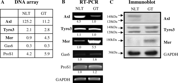
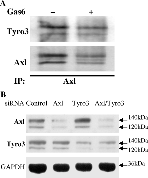
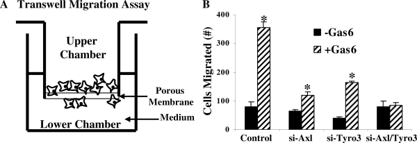
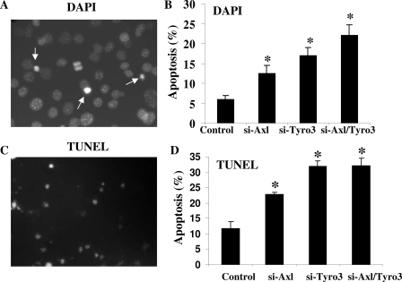
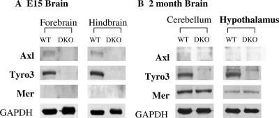
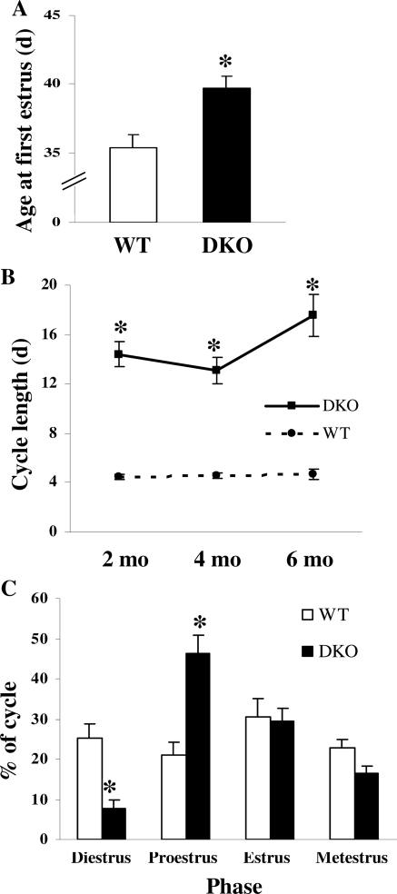
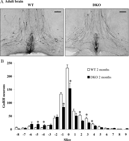
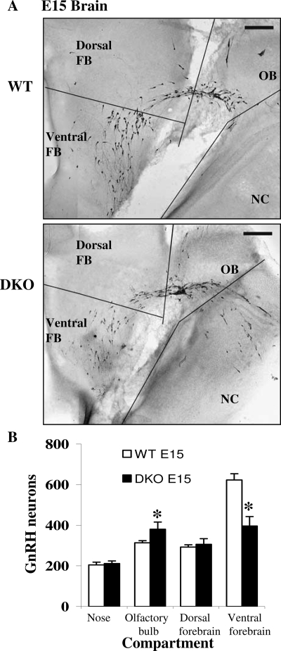
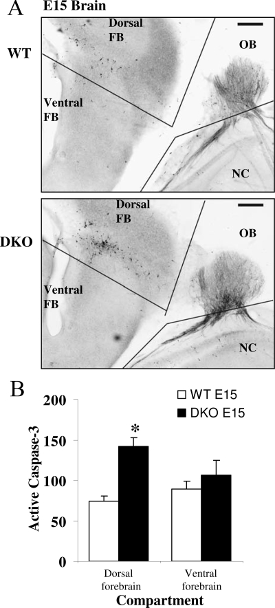
GnRH neurons must undergo a complex and precise pattern of neuronal migration to appropriately target their projections to the median eminence to trigger gonadotropin secretion and thereby control reproduction. Using NLT GnRH cells as a model of early GnRH neuronal development, we identified the potential importance of Axl and Tyro3, members of the TAM (Tyro3, Axl, and Mer) family of receptor tyrosine kinases in GnRH neuronal cell survival and migration. Silencing studies evaluated the role of Tyro3 and Axl in NLT GnRH neuronal cells and suggest that both play a role in Gas6 stimulation of GnRH neuronal survival and migration. Analysis of mice null for both Axl and Tyro3 showed normal onset of vaginal opening but delayed first estrus and persistently abnormal estrous cyclicity compared with wild-type controls. Analysis of GnRH neuronal numbers and positioning in the adult revealed a total loss of 24% of the neuronal network that was more striking (34%) when considered within specific anatomical compartments, with the largest deficit surrounding the organum vasculosum of the lamina terminalis. Analysis of GnRH neurons during embryogenesis identified a striking loss of immunoreactive cells within the context of the ventral forebrain compartment (36%) and not more rostrally. Studies using caspase 3 cleavage as a marker of apoptosis showed that Axl(-/-), Tyro3(-/-) double-knockout mice had increased cell death in the nose and dorsal forebrain, supporting the underlying mechanism of cell loss. Together these data suggest that Axl and Tyro3 mediate the survival and appropriate targeting of GnRH neurons to the ventral forebrain, thereby contributing to normal reproductive function and cyclicity in the female.


Figures

Figure 1
Figure 1
Differential Expression of TAM Family Members in GnRH Neuronal Cells A, Normalized transcript levels of Axl, Tyro3, Mer, Gas6, and ProS1 from triplicate DNA microarray analysis (see Materials and Methods for details). B, Semiquantitative RT-PCR of TAM family member mRNA levels in NLT and GT1-7 GnRH neuronal cells. Numbers refer to fold differences in TAM mRNA levels between NLT and GT1-7 cells. C, Immunoblot of TAM family member protein levels expressed in NLT and GT1-7 GnRH neuronal cells. GAPDH was used as a loading control.
Figure 2
Figure 2
Dissecting the Individual Roles of Axl and Tyro3 in GnRH Neuronal Cells A, Tyro3 and Axl form heterodimers in GnRH neuronal cells. After 10 min incubation in the absence or presence of 400 ng/ml Gas6, NLT lysates were immunoprecipitated with anti-Axl and then immunoblotted with anti-Axl or anti-Tyro3. B, siRNAs inhibit Axl and Tyro3 in GnRH neuronal cells. NLT cells were transfected with control scrambled siRNA or siRNA specific for Axl, Tyro3, or both. Twenty-four hours after transfection, cells were harvested for immunoblot and probed with antibodies for Axl, Tyro3, and GAPDH (as a loading control).
Figure 3
Figure 3
Contribution of Axl and Tyro3 to Gas6-Mediated Activation of Migration in NLT GnRH Neuronal Cells A, Schematic presentation of transwell migration assay. B, Functional effects of silencing Axl, Tyro3, or both on Gas6-induced NLT GnRH neuronal migration (see Materials and Methods for details). *, P < 0.05; n = 3.
Figure 4
Figure 4
Effects of Silencing Axl and Tyro3 to Rates of Programmed Cell Death in GnRH Neuronal Cells Photomicrographs of NLT neurons assessed for DAPI staining (A) or TUNEL (C) to detect apoptotic nuclei. Summary of the functional effects of silencing Axl, Tyro3, or both on rates of apoptosis in response to serum deprivation as assessed by DAPI (B) or TUNEL (D) (see Materials and Methods for details). *, P < 0.05; n = 3.
Figure 5
Figure 5
Expression of TAM Family Member Protein Levels in Brains of E15 and Adult WT and DKO Mice A, Expression of Axl, Tyro3, and Mer in the forebrain and hindbrain regions of E15 WT but not DKO mice. B, Differential expression of Axl, Tyro3, and Mer in brain regions from WT and DKO mice at 2 months of age. See Materials and Methods for details.
Figure 6
Figure 6
Axl and Tyro3 Impact on the Process of Sexual Maturation in Female Mice A, DKO mice (n = 17) have delayed first estrus compared with WT controls (n = 10). *, P < 0.05. B, DKO mice have prolonged irregular estrous cycle length at 2, 4, and 6 months. *, P < 0.05 (n = 10 for DKO time points; n = 4 for WT time points). C, DKO mice (n = 10) have increased proestrous and decreased diestrous phase lengths compared with WT mice (n = 4) at 2 months. *, P < 0.05.
Figure 7
Figure 7
Two-Month-Old Axl/Tyro3-Null Mice Have Decreased Numbers of GnRH Neurons in the Hypothalamus A, Images of immunoreactive GnRH neurons in WT (n = 4) and DKO (n = 5) mice taken at ×200 with the OVLT shown in the bottom center of each image. B, Brains from DKO mice had fewer immunoreactive GnRH neurons compared with WT in slices −1, 0, and +2–3 but more in slices −5 and −4. GnRH neuron counts were obtained in slices that represent 330 μm each, with OVLT labeled as zero (see Materials and Methods for details). *, P < 0.05.
Figure 8
Figure 8
GnRH Neuronal Number and Position Are Altered in E15 DKO Brains A, Low-magnification images of immunoreactive GnRH neurons in WT and DKO E15 heads showing the locations of the compartments that were counted. FB, Forebrain; NC, nasal compartment; OB, olfactory bulb. B, Altered distribution of GnRH neurons in separate compartments of E15 DKO brains (n = 5 for both genotypes). *, P < 0.05.
Figure 9
Figure 9
Apoptosis in Vivo in DKO Compared with WT Embryonic Mice Photomicrographs of sagittal sections of E15 brain showing cleaved caspase 3 immunoreactivity in WT (A) and DKO (B). C, Results of analysis of active caspase 3 cleavage in counts of cells in dorsal and ventral forebrain of WT (white bars) and DKO (black bars). n = 5; *, P < 0.05 from WT control.

Similar articles

Cited by

References

    1. Wray S, Hoffman G 1986 Postnatal morphological changes in rat LHRH neurons correlated with sexual maturation. Neuroendocrinology 43:93–97 - PubMed
    1. Schwanzel-Fukuda M, Bick D, Pfaff DW 1989 Luteinizing hormone-releasing hormone (LHRH)-expressing cells do not migrate normally in an inherited hypogonadal (Kallmann) syndrome. Brain Res Mol Brain Res 6:311–326 - PubMed
    1. Yoshida K, Tobet SA, Crandall JE, Jimenez TP, Schwarting GA 1995 The migration of luteinizing hormone-releasing hormone neurons in the developing rat is associated with a transient, caudal projection of the vomeronasal nerve. J Neurosci 15:7769–7777 - PMC - PubMed
    1. Tobet SA, Schwarting GA 2006 Recent progress in gonadotropin-releasing hormone neuronal migration. Endocrinology 147:1159–1165 - PubMed
    1. Schwarting GA, Wierman ME, Tobet SA 2007 Gonadotropin-releasing hormone neuronal migration. Semin Reprod Med 25:305–312 - PubMed

Publication types

MeSH terms

Substances