. 2008 Oct;173(4):1120-8.
doi: 10.2353/ajpath.2008.080252. Epub 2008 Sep 11.

Gastrointestinal dysmotility in mitochondrial neurogastrointestinal encephalomyopathy is caused by mitochondrial DNA depletion

Affiliations

Gastrointestinal dysmotility in mitochondrial neurogastrointestinal encephalomyopathy is caused by mitochondrial DNA depletion

Carla Giordano et al. Am J Pathol. 2008 Oct.

Abstract

Chronic intestinal pseudo-obstruction is a life-threatening condition of unknown pathogenic mechanisms. Chronic intestinal pseudo-obstruction can be a feature of mitochondrial disorders, such as mitochondrial neurogastrointestinal encephalomyopathy (MNGIE), a rare autosomal-recessive syndrome, resulting from mutations in the thymidine phosphorylase gene. MNGIE patients show elevated circulating levels of thymidine and deoxyuridine, and accumulate somatic mitochondrial DNA (mtDNA) defects. The present study aimed to clarify the molecular basis of chronic intestinal pseudo-obstruction in MNGIE. Using laser capture microdissection, we correlated the histopathological features with mtDNA defects in different tissues from the gastrointestinal wall of five MNGIE and ten control patients. We found mtDNA depletion, mitochondrial proliferation, and smooth cell atrophy in the external layer of the muscularis propria, in the stomach and in the small intestine of MNGIE patients. In controls, the lowest amounts of mtDNA were present at the same sites, as compared with other layers of the gastrointestinal wall. We also observed mitochondrial proliferation and mtDNA depletion in small vessel endothelial and smooth muscle cells. Thus, visceral mitochondrial myopathy likely causes gastrointestinal dysmotility in MNGIE patients. The low baseline abundance of mtDNA molecules may predispose smooth muscle cells of the muscularis propria external layer to the toxic effects of thymidine and deoxyuridine, and exposure to high circulating levels of nucleosides may account for the mtDNA depletion observed in the small vessel wall.


Figures
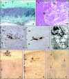
Figure 1
Figure 1
Morphological features of the GI tract in MNGIE. A: The wall of small intestine shows marked atrophy and fibrosis of the external layer (EL) of muscularis propria. Few residual smooth muscle cells are evident within the fibrous tissue (arrows). The internal layer (IL) is unremarkable (patient 4, Masson trichrome stain, original magnification ×10). B: Residual smooth muscle cells surrounded by fibrous tissue in the external layer (EL) of muscularis propria show extensive vacuolation and pyknotic nuclei. A morphologically normal myenteric plexus is marked by the asterisk. (patient 1, H&E stain, original magnification ×20). C: Immunostain for mitochondrial antigens shows marked mitochondrial proliferation in smooth muscle cells of internal (IL) and external (EL) layers of muscularis propria from small intestine of MNGIE patient 2 (C) as compared with one control (D). Of note, the density of mitochondria in ganglion cells (asterisk) is high as compared with smooth muscle cells of the internal (IL) and external (EL) layer of tunica muscularis (anti-mitochondrial antigens antibody, clone MTC, UCS Diagnostic, original magnification ×10). E: Ultrastructural features of smooth muscle cells in the small intestine of MNGIE patient 1. Smooth muscle cells are identified by the presence of “focal adhesions” (square boxes). The cell cytoplasm shows numerous mitochondria (arrows) with postmortem artifactual swelling. F: The combined COX/SDH stain shows numerous COX-negative smooth muscle cells in the internal layer (IL) of muscularis propria of small intestine. These cells appear blue since they are devoid of the COX activity (orange) but retain the SDH activity (blue). Two blue COX-negative ganglion cells are marked by the asterisk (patient 3, COX/SDH, original magnification ×10). G: Combined COX/SDH stain in the small intestinal wall of a control subject. Note the absence of blue COX-negative muscle fibers Ganglion cells stained with COX are marked by an asterisk (IL, internal layer; EL, external layer; COX/SDH, original magnification ×10). H: The combined COX/SDH stain of the muscularis propria of small intestine shows patchy areas with COX-negative smooth muscle cells in blue marked by arrows. (patient 3, COX/SDH, original magnification ×20).
Figure 2
Figure 2
Mitochondrial proliferation in smooth muscle and endothelial cells of small vessels in MNGIE. A: Immunostain for mitochondrial antigens shows mitochondrial proliferation in the tunica media of a small artery from a MNGIE patient (patient 1, anti-mitochondrial antigens antibody, clone MTC, UCS Diagnostic, original magnification ×10). B: Immunostain for mitochondrial antigens in a small artery from a control subject (Anti-mitochondrial antigens antibody, clone MTC, UCS Diagnostic, original magnification ×10). C: The combined COX/SDH stain shows a blue COX-negative vessel from the small bowel surrounded by brown COX-positive smooth muscle cells of muscularis propria (patient 3, COX/SDH, original magnification ×40). D: Ultrastructural features of endothelial cells from a small vessel of MNGIE patient 2. Note the numerous mitochondria (arrows).with postmortem artifactual swelling. A red blood cell is marked by the asterisk.
Figure 3
Figure 3
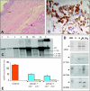
Histopathology and molecular analysis of esophagus in MNGIE. A: The wall of proximal esophagus has a normal appearance. The arrow indicates smooth muscle cells of tunica muscularis and the asterisk marks the striated fibers of the cricopharyngeal muscle (patient 2, H&E, original magnification ×4). B: The combined COX/SDH stain shows frequent COX-negative fibers in blue (patient 3, COX/SDH, original magnification ×40). C: Southern blot analysis of DNA from tissue homogenates of patient 1 shows multiple deletions only in the esophagus and skeletal muscle. In addition to the signal from the 16.6-kb mtDNA molecule (WT), signals of lower molecular weight, corresponding to mtDNA deleted molecules (arrows), are evident. C, colon; S, stomach; I, ileum; E, esophagus; and M, skeletal muscle. M+ is a positive control with multiple deletion. M− is a control subject. U, uncut sample. D: The Δ5-Kb, Δ7.7-Kb, Δ8.1-Kb, and Δ9.5-Kb deletions were investigated by PCR assays using oligonucleotide primers flanking the regions upstream and downstream breakpoints in the parental molecule (see Materials and Methods). Representative 2% agarose gels from patient 3. The bands corresponding to the mtDNA deletions were detected in esophagus homogenate (E) and in the microdissected striated fibers from cricopharyngeal muscle (Sk). No deletions were detected in smooth muscle cells from tunica muscularis (Sm). (Negative control, C−; the arrow indicate 500 bp). E: MtDNA content was evaluated in single COX-positive (blue bar) and COX-negative (green bar) skeletal muscle fibers from cricopharyngeal muscle from MNGIE patients 3 and 4 versus three controls (red bar). In MNGIE patients, mtDNA/nuclear DNA ratio is less than normal controls, in both COX-positive and COX-negative fibers. Severely COX-deficient fibers have lower mtDNA amount than fibers with residual COX activity.
Figure 4
Figure 4
Assessment of mtDNA content in the different GI segments. A: Interaction plot of mtDNA copies/cells means (after transformation to the natural logarithm) ± 2 standard errors (SE) versus GI tissue segment homogenates for patients (red line) and controls (black lines). MNGIE patients shows a striking reduction in mtDNA content limited to the small intestine (P < 0.001 vs controls). A milder, although significant, decrease is observed in the stomach (P < 0.01 vs controls). In control subjects, the small intestine has a lower amount of mtDNA relative to esophagus, stomach, and colon. B: Interaction plot of mtDNA copies/cells means (after transformation to the natural logarithm) ± 2 SE versus microdissected tissues of stomach, small intestine and colon, for MNGIE patients (green line) and controls (red line). In MNGIE patients the external (EL) layers of muscularis propria from all segments of the GI wall show significant reductions in mtDNA content as compare to controls (P < 0.001). The lowest mtDNA content is observed in the EL of small intestine (93% decrease). A milder although significant decrease is observed in the internal layer (IL) from the entire GI tract and in the myenteric plexus (MP) of small intestine (P < 0.001). In control subjects, the EL of muscularis propria shows a lower amount of mtDNA as compared to the IL (internal/external layer ratio 2:1). C: Regression analysis between mtDNA copies/cells means in different microdissected tissues from MNGIE patients and controls. It is shown a linear relationship between the mtDNA amount in different tissue components of GI wall, (ie, internal and external layer of muscularis propria and myenteric plexus from stomach, small intestine, and colon) from MNGIE patients and control subjects (black dots) (r2 = 0.89; P < 0.0001). In contrast, microdissected small vessels (red dot) show marked mtDNA depletion in MNGIE patients, despite the high mtDNA levels in controls. S = stomach; I = small intestine; C = colon; 1 = myenteric plexus; 2 = internal, and 3 = external, layers of muscularis propria.

Similar articles

Cited by

References

    1. Connor FL, Di Lorenzo C. Chronic intestinal pseudo-obstruction: assessment and management. Gastroenterology. 2006;30:S29–S36. - PubMed
    1. Stanghellini V, Cogliandro RF, De Giorgio R, Barbara G, Salvioli B, Corinaldesi R. Chronic intestinal pseudo-obstruction: manifestations, natural history and management. Neurogastroenterol Motil. 2007;19:440–452. - PubMed
    1. De Giorgio R, Sarnelli G, Corinaldesi R, Stanghellini V. Advances in our understanding of the pathology of chronic intestinal pseudo-obstruction. Gut. 2004;53:1549–1552. - PMC - PubMed
    1. Jain D, Moussa K, Tandon M, Culpepper-Morgan J, Proctor DD. Role of interstitial cells of Cajal in motility disorders of the bowel. Am J Gastroenterol. 2003;98:618–624. - PubMed
    1. Bindoff L. Mitochondrial gastroenterology. Di Mauro S, Hirano M, Schon EA, editors. Abington, UK: Informa Healthcare,; Mitochondrial Medicine. 2006:143–159.

Publication types

MeSH terms

Substances