. 2008 Oct;173(4):1229-42.
doi: 10.2353/ajpath.2008.080014. Epub 2008 Sep 11.

Nuclear interleukin-33 is generally expressed in resting endothelium but rapidly lost upon angiogenic or proinflammatory activation

Affiliations

Nuclear interleukin-33 is generally expressed in resting endothelium but rapidly lost upon angiogenic or proinflammatory activation

Axel M Küchler et al. Am J Pathol. 2008 Oct.

Abstract

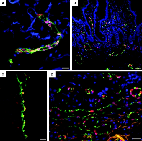
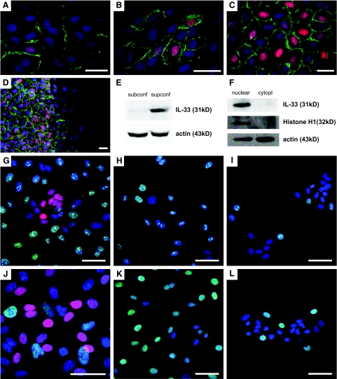
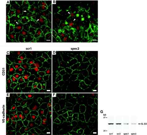
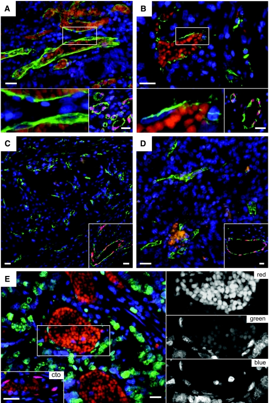
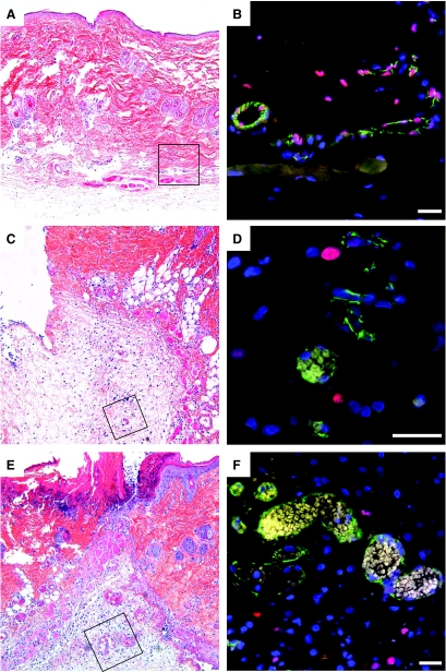
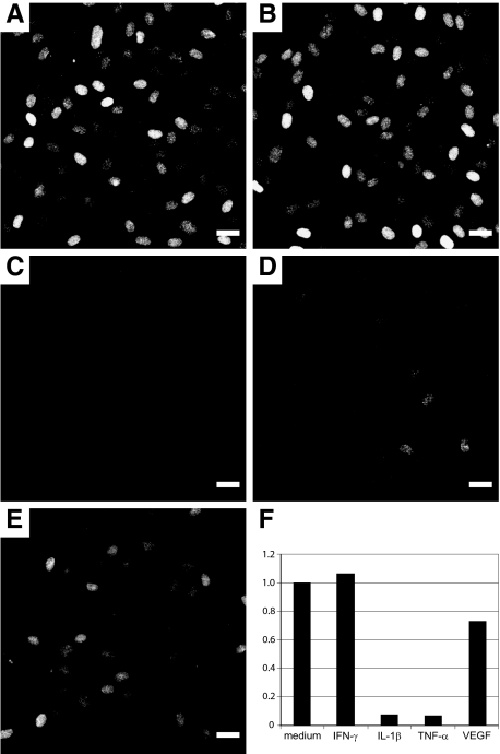
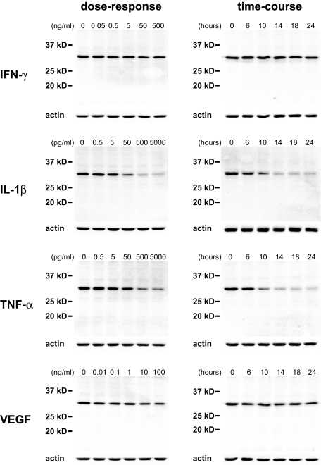
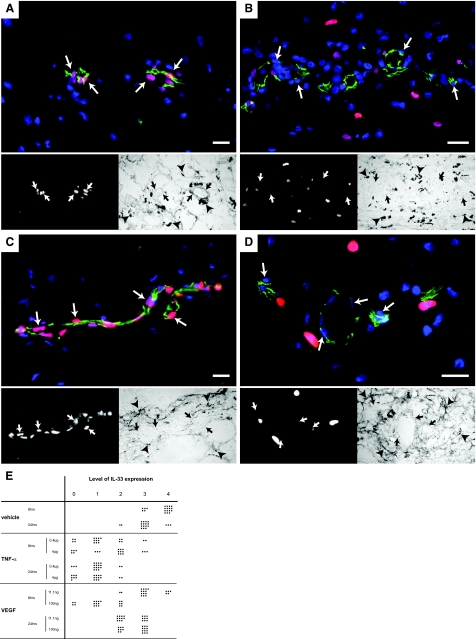
Interleukin (IL)-33 is a novel member of the IL-1 family of cytokines that promotes Th2 responses in lymphocytes as well as the activation of both mast cells and eosinophils via the ST2 receptor. Additionally, IL-33 has been proposed to act as a chromatin-associated transcriptional regulator in both endothelial cells of high endothelial venules and chronically inflamed vessels. Here we show that nuclear IL-33 is expressed in blood vessels of healthy tissues but down-regulated at the earliest onset of angiogenesis during wound healing; in addition, it is almost undetectable in human tumor vessels. Accordingly, IL-33 is induced when cultured endothelial cells reach confluence and stop proliferating but is lost when these cells begin to migrate. However, IL-33 expression was not induced by inhibiting cell cycle progression in subconfluent cultures and was not prevented by antibody-mediated inhibition of VE-cadherin. Conversely, IL-33 knockdown did not induce detectable changes in either expression levels or the cellular distribution of either VE-cadherin or CD31. However, activation of endothelial cell cultures with either tumor necrosis factor-alpha or vascular endothelial growth factor and subcutaneous injection of these cytokines led to a down-regulation of vascular IL-33, a response consistent with both its rapid down-regulation in wound healing and loss in tumor endothelium. In conclusion, we speculate that the proposed transcriptional repressor function of IL-33 may be involved in the control of endothelial cell activation.


Figures

Figure 1
Figure 1
IL-33 is globally expressed in nuclei of vascular endothelium in normal human tissues. Endothelial cells in vessels of human skin (A), small intestine (B), umbilical vein (C), and lung (D) were stained for IL-33 (Nessy-1, red) and with Ulex europaeus agglutinin I (A, C, and D; green) or CD31 (B, green) and Hoechst dye (blue) to visualize endothelial cells and cell nuclei, respectively. Three-color fluorescence micrograph of fixed and permeabilized tissue sections are shown. Note of presence of IL-33 positive single tissue resident cells in (B) and (C). Scale bars = 50 μm.
Figure 2
Figure 2
IL-33 in HUVECs responds to cell density signals and is localized to the nucleus in nonproliferating, superconfluent cells. A–D: Monolayers of HUVECs stained for IL-33 (red) using IL-33Nterm (A–C) and Nessy-1 (D). CD31 is stained in green and cell nuclei are visualized by Hoechst dye (blue). A and B: HUVECs induce expression of nuclear IL-33 and enhance the localization of CD31 to the intercellular areas when cell density increases. C: Almost all cells are IL-33-positive in a superconfluent monolayer. D: Superconfluent HUVEC monolayer 4 hours after scratch wounding. The red signal in the cytoplasm cannot be distinguished from the signal obtained with irrelevant antibodies (data not shown). Note loss of nuclear IL-33 and reduction of CD31 (green) in cells that migrate into the open area (right half of image). E and F: Western blot for IL-33 detection in whole cell lysates of subconfluent (subconf) or superconfluent (supconf) cells (E) or nuclear versus cytoplasmic (cytopl) fraction of a superconfluent HUVEC monolayer (F). Monolayers of HUVECs stained for IL-33 (IL-33Nter in G, Nessy-1 in H–L; all red), Ki-67 (G--I, green), and ppRb (J–L, green). Endothelial nuclei are visualized by Hoechst dye (blue). Note the lack of IL-33 expression in serum and growth factor starved cells in I and L. Scale bars: 25 μm (A–D); 30 μm (G–L).
Figure 3
Figure 3
Inhibition of the junctional protein VE-cadherin does not alter the expression level of IL-33 and knockdown of IL-33 does not alter the cellular distribution or expression level of junctional proteins CD31 or VE-cadherin. Immunocytochemical double staining for IL-33 (IL-33Nter, red) and VE-cadherin (mouse IgG1, green) in HUVECs preincubated with vehicle (PBS, A) or anti-VE-cadherin (IgG2a, B) from point of seeding until fixation. Pictures were obtained using identical exposure times and image-enhancement parameters. Arrowheads point to areas of intercellular contact. Note that cells in B with redistribution or lowered expression level of VE-cadherin maintained expression of IL-33. C and D: Immunocytochemical double staining for IL-33 (IL-33Nter, red) and CD31 (green) in HUVECs transfected with mock (C) or specific siRNA (D). E and F: Immunocytochemical double staining for IL-33 (red) and VE-cadherin (green) in HUVECs transfected with mock (E) or specific siRNA (F). Pictures were obtained using identical exposure times and image enhancement parameters. G: Western blot of IL-33 in cell lysates of HUVEC monolayers transfected with either scrambled siRNA controls (scr1 and scr2) or concentration-matched IL-33 targeting siRNA (spec1 and spec2). Shown are lanes from the same experiment, which was independently repeated twice with similar results. Scale bars = 10 μm.
Figure 4
Figure 4
Vascular IL-33 expression is lost in tumor vessels. Endothelial cells in vessels of human colon adenocarcinoma (A), ductal mammary carcinoma (B), renal clear cell carcinoma (C), and renal papillary carcinoma (D) were stained for IL-33 (Nessy-1, red) and with Ulex europaeus lectin (green) and Hoechst dye (blue) to visualize endothelial cells and cell nuclei, respectively. (For consistency throughout the figures, the red and green channels in Figure 4 were reversed.) The bottom left panels in A and B show high-power magnifications of the respective boxed areas. Note presence of autofluorescent intravascular red blood cells in A and B as well as lack of IL-33 signal in endothelial cell nuclei. The bottom right panels in A to D and bottom left panel in E show IL-33-positive vessels in tissue surrounding the tumor in the same section. E: Human colon adenocarcinoma (different sample from A) was stained for IL-33 (Nessy-1, red), Ki-67 (green), and Hoechst dye (blue). Autofluorescent erythrocytes indicate blood vessels. Right: High-power magnifications of the boxed area in E separating the different colors. Note the lack of IL-33 signal in Ki-67-positive endothelial nuclei. Scale bars = 50 μm.
Figure 5
Figure 5
Vascular IL-33 expression is lost during wound healing. Biopsies from normal rat skin (A and B) and from wounds after 1 (C and D) or 2 days (E and F) were fixed and stained with H&E (A, C, and E) or immunostained (B, D, and F) for IL-33 (Nessy-1, red) and CD31 (green). A, C, and E show overviews of the wound area and the boxed areas indicate the position of vessels shown in B, D, and F, respectively. Cell nuclei are stained with Hoechst dye. Note presence of vessels without IL-33 and perivascular cell nuclei at days 1 and 2. Scale bars = 50 μm.
Figure 6
Figure 6
IL-33 expression is down-regulated by proinflammatory cytokines and VEGF. A--E: Response of superconfluent HUVEC monolayers exposed for 17 hours to medium alone (A), INF-γ (50 ng/ml) (B), IL-1β (0.5 ng/ml) (C), TNF-α (0.5 ng/ml) (D), or VEGF (10 ng/ml) (E). Immunostaining for IL-33 (IL-33Nter) after fixation of monolayers. Pictures were obtained using identical exposure times and image enhancement parameters. F: Quantitative PCR analysis of IL-33 expression in superconfluent HUVEC monolayers treated with cytokines for 7 hours at the same concentrations as given above. IL-33 transcript levels were obtained using primer pair IL-33 no. 2 and normalized to HPRT, data are given as ratio of transcript values in sample over those in medium-treated controls. Similar results were obtained when using a different primer pair for IL-33 detection or when normalizing against housekeeping gene GAPDH (data not shown). Scale bars = 25 μm.
Figure 7
Figure 7
Dose-response and time-course of IL-33 expression in HUVECs. Western blot detection of IL-33 protein after cytokine treatment of superconfluent HUVEC monolayers for 18 hours with the indicated concentration of cytokines (left column) or for the indicated time with INF-γ (50 ng/ml), IL-1β (500 pg/ml), TNF-α (500 pg/ml), or VEGF (10 ng/ml) (right column). Note the sensitivity of IL-33 levels to IL-1β and TNF-α whereas VEGF has a mild effect.
Figure 8
Figure 8
TNF-α and VEGF down-regulate IL-33 in vivo. Immunostaining for IL-33 (Nessy-1, red) and CD31 (green) of rat skin after subcutaneous injection of PBS (A, C) and cytokines (B, D). Cell nuclei are stained with Hoechst dye. The bottom left panels show the red channel and the bottom right panels the light microscopy images of the respective area. Twenty-four hours after the injection of 0.4 μg of rrTNF-α (B) or 8 hours after the injection of 100 ng of rrVEGF (D) the endothelial signal for IL-33 has decreased compared to the mock injection (A and C, respectively). Pictures were obtained using identical exposure times and image enhancement parameters for A and B as well as for C and D. Arrows indicate endothelial nuclei. Arrowheads indicate ink particles. E: Semiquantitative analysis of vascular IL-33 expression in rat skin after injection of vehicle (PBS), rrTNF-α, or rrVEGF. Endothelial cell nuclei were categorized as negative (0) or increasingly positive (levels 1 to 4) by a blinded observer, counting in each condition 20 nuclei. Scale bars = 50 μm.

References

    1. Onda H, Kasuya H, Takakura K, Hori T, Imaizumi T, Takeuchi T, Inoue I, Takeda J. Identification of genes differentially expressed in canine vasospastic cerebral arteries after subarachnoid hemorrhage. J Cereb Blood Flow Metab. 1999;19:1279–1288. - PubMed
    1. Baekkevold ES, Roussigne M, Yamanaka T, Johansen FE, Jahnsen FL, Amalric F, Brandtzaeg P, Erard M, Haraldsen G, Girard JP. Molecular characterization of NF-HEV, a nuclear factor preferentially expressed in human high endothelial venules. Am J Pathol. 2003;163:69–79. - PMC - PubMed
    1. Schmitz J, Owyang A, Oldham E, Song Y, Murphy E, McClanahan TK, Zurawski G, Moshrefi M, Qin J, Li X, Gorman DM, Bazan JF, Kastelein RA. IL-33, an interleukin-1-like cytokine that signals via the IL-1 receptor-related protein ST2 and induces T helper type 2-associated cytokines. Immunity. 2005;23:479–490. - PubMed
    1. Carriere V, Roussel L, Ortega N, Lacorre DA, Americh L, Aguilar L, Bouche G, Girard JP. IL-33, the IL-1-like cytokine ligand for ST2 receptor, is a chromatin-associated nuclear factor in vivo. Proc Natl Acad Sci USA. 2007;104:282–287. - PMC - PubMed
    1. Sanada S, Hakuno D, Higgins LJ, Schreiter ER, McKenzie AN, Lee RT. IL-33 and ST2 comprise a critical biomechanically induced and cardioprotective signaling system. J Clin Invest. 2007;117:1538–1549. - PMC - PubMed

Publication types

MeSH terms