. 2008 Oct;173(4):927-37.
doi: 10.2353/ajpath.2008.071149. Epub 2008 Sep 11.

Deletion of CD151 results in a strain-dependent glomerular disease due to severe alterations of the glomerular basement membrane

Affiliations

Deletion of CD151 results in a strain-dependent glomerular disease due to severe alterations of the glomerular basement membrane

Rosa M Baleato et al. Am J Pathol. 2008 Oct.

Abstract

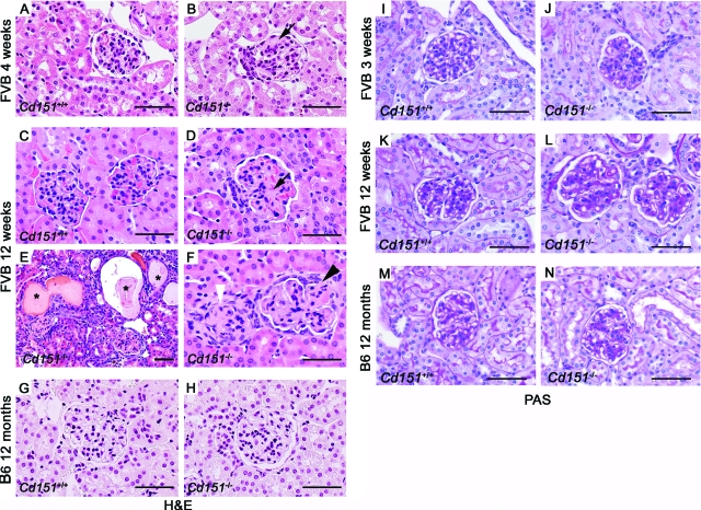
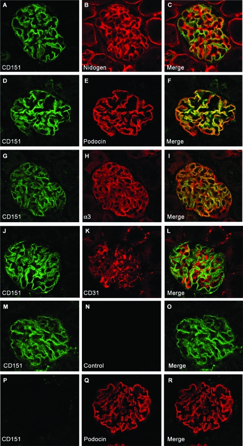
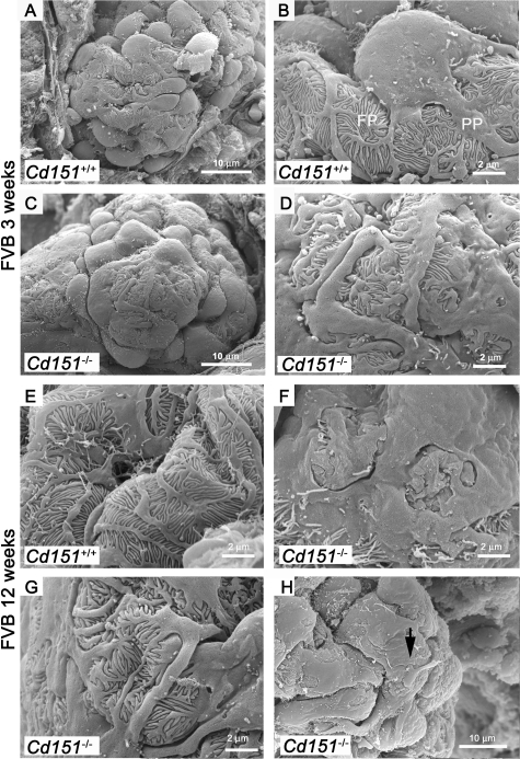
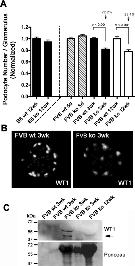
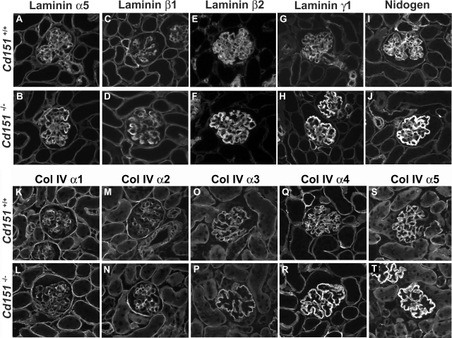
Alterations in CD151 have been associated with primary glomerular disease in both humans and mice, implicating CD151 as a key component of the glomerular filtration barrier. CD151 belongs to the tetraspanin family and associates with cell-matrix adhesion complexes such as alpha3beta1-integrin. Here we show that Cd151-deficient mice develop severe kidney disease on an FVB background but are healthy on a B6 background, providing a new and unique tool for the identification of genes that modulate the onset of proteinuria. To better understand the function of CD151 in the kidney, we studied its expression pattern and characterized early ultrastructural defects in Cd151-null kidneys. CD151 is expressed in podocytes of the mouse kidney and co-localizes with alpha3-integrin at the base of podocyte foot processes, at the site of anchorage to the glomerular basement membrane (GBM). Interestingly, the first ultrastructural lesions seen at the onset of proteinuria in Cd151-null kidneys were severe alterations of the GBM, reminiscent of Alport syndrome and consisting of massive thickening and splitting of the GBM. These lesions are associated with increased expression of GBM components. Podocyte abnormalities, effacement of foot processes, and podocyte loss appear to occur consequently to the GBM damage. In conclusion, CD151 appears to be involved in the establishment, maturation, and/or maintenance of the GBM structure in addition to its role in integrin-mediated adhesion strengthening.


Figures

Figure 1
Figure 1
Massive Proteinuria in Cd151-Null Mice on the FVB but Not B6 Background. Urine from Cd151+/+ and Cd151−/− mice at different ages was analyzed by SDS- polyacrylamide gel electrophoresis. One microliter of urine from each mouse was loaded on the same gel. Proteinuria was observed as early as 5 days of age in the FVB Cd151-null mouse and increased substantially with age to become massive at 12 weeks (note that the urine sample of the 12 week-old FVB Cd151-null mouse has been diluted 1:10). B6 Cd151-null mice were analyzed up to 12 months of age and did not exhibit any signs of proteinuria.
Figure 2
Figure 2
Histological Analysis of Cd151−/− Kidneys. Cd151+/+ and Cd151−/− Kidneys from both FVB (A–F; I–L) and B6 strains (G, H, M, N) were analyzed by H&E (A–H) and PAS staining (I–N). At 4 weeks of age, FVB Cd151-null mice presented with mild glomerular damage as seen by H&E staining (black arrow head in B) compared to wild-type (A). At this age, the glomerular damage consisted exclusively of GBM thickening as seen on the PAS stains (I, J). By 12 week of age, all FVB Cd151-null mice showed significantly increased kidney damage (D–F) compared to wild-type control (C), ranging from moderate glomerular damage (D, black arrow) to more severe glomerular damage associated with tubular lesions (E). The GBM of Cd151−/− mice at this age was extensively thickened (L). In addition, a few 12-week-old FVB Cd151-null mice presented with massive glomerulosclerosis and collapse of the glomerular tuft (E, F, white arrowhead) associated with massive tubular dilations and protein casts (E*). Some sclerotic glomeruli showed partial (F, black arrowhead) or complete (F, white arrowhead) adhesion to the Bowman’s capsule. The 12 month-old B6 Cd151-null kidneys appeared totally normal (H, N) as compared with wild-type controls (G, M). Scale bars = 50 μm.
Figure 3
Figure 3
Localization of CD151 in the Mouse Kidney. Dual immunofluorescence labeling and confocal analysis using CD151 antibody (green) and antibodies (red) directed against nidogen (A–C), podocin (D–F), α3 integrin (G–I), and CD31 (J–L). The CD151 antibody labeled mainly glomeruli in the kidney (A–O). The specificity of the CD151 antibody was shown by absence of CD151 labeling on Cd151−/− section (P–R). The green CD151 labeling follows the GBM (red, C) and colocalizes substantially with podocin (F) and α3 integrin (I) at the base of podocytes. CD151 does not colocalize with CD31 in the glomerular endothelium (L). M–O: For labeling where two rabbit antibodies were used, an appropriate control (where the second primary antibody was omitted) was included in the experiments (M–O). The absence of labeling by the second secondary antibody (red) in this control proves sufficient blocking of the first primary (CD151) and secondary antibody (green). Pictures were taken using a confocal microscope. Original magnification ×400.
Figure 4
Figure 4
TEM study of FVB Cd151−/− Kidneys. Five-day-old (A–D) and 3-week-old (E–H) FVB kidneys were analyzed by TEM. The GBM is abnormally and irregularly thickened in the 5-day- and 3-week-old FVB Cd151-null animals (*in B, C, F, and G). Normal differentiation and architecture of the glomerular filtration barrier in FVB wild-type animals is shown at 5 days (A) and 3 weeks (E) of age. GBM maturation is not yet complete in the 5-day-old wild-type FVB mice (box in A, white arrowhead). The striking feature in Cd151-null kidneys is the massive thickening and splitting of the GBM observed as early as 5 days in some segments of glomeruli (B, C*). The GBM disorganization becomes more important and involves more capillary loops with progression of the disease as shown here at 3 weeks of age (F, G*). Foot processes and slit diaphragms seem to develop normally as observed in the 5-day-old (C, D, arrows) and 3-week-old (H, arrow) Cd151−/− animals (C, D arrows). The foot processes were effaced in a few areas (arrowhead in B and G) but they were surprisingly well maintained in other areas, given the extent of GBM damage (C, D, and H). A semiquantitative analysis of the extent of GBM splitting (I) and effacement of foot processes (J) over time was performed. The results are expressed as percentage of split GBM and number of podocyte foot processes (FP) per micrometer of GBM, respectively. The representative results from two mice in each group at 5 days and 3 weeks are shown. Scale bars = 2 μm (A, B, E, F); 500 nm C, D, G, H.
Figure 5
Figure 5
SEM Analysis of Podocyte Structure in FVB Cd151-Null Mice. Three-and 12-week old FVB Cd151+/+ and Cd151−/− kidneys were analyzed by SEM to determine the extent of glomerular and more specifically podocyte damage. In wild-type animals (A, B, and E) the podocyte structure is well defined with the cell body dividing into primary processes (PP) that divide into foot processes (FP). At 3 weeks of age, the podocytes look abnormal in Cd151−/− kidneys (C, D) compared to wild-type controls (A, B). Indeed, the podocyte cell bodies look flattened at 3 weeks of age (C). Primary processes and foot processes are still present in most areas as seen by TEM but appear disorganized (C, D, and G). Podocyte alterations progress with age and foot process effacement has become more pronounced by 12 weeks (F). However, even at 12 weeks of age, there are still a number of places where disorganized foot processes are still visible (G). Also note the unhealthy podocyte sending a process onto the cell body of its neighbor (arrow in H). The representative results from 3 mice in each group at 3 weeks of age and two mice in each group at 12 weeks of age are shown. Three to 6 glomeruli were analyzed for each mouse. Original magnification: ×6000 (A, C); ×20,000 (B, D–G); ×12,000 (H).
Figure 6
Figure 6
Progressive Podocyte Loss in FVB Cd151−/− Kidneys. A: Podocyte number per glomerulus was estimated by counting nuclei positive for both WT1 and DAPI stainings. Results were normalized to the average podocyte number in the wild-type control for each age. At 5 days of age podocyte number in Cd151−/− kidneys was equal to control, showing that the podocytes differentiate normally. At 3 weeks and 12 weeks of age FVB Cd151-deficient mice present with a significant decrease in podocyte number per glomerulus (22.2% and 28.4%, respectively), compared to wild-type controls (P < 0.001). In B6 Cd151-null animals there is no loss in podocyte number compared to wild-type controls. B: Representative pictures of the WT1 immunolabeling used to assess the number of podocytes per glomerulus in both wild-type (wt) and Cd151 knock-out (ko) kidney sections. C: Representative Western blot of urine from Cd151+/+ and Cd151−/− mice probed with WT1 antibody (top panel), showing presence of podocytes in urine of some Cd151−/− mice at 3 weeks and 12 weeks of age (black arrow). Ponceau staining of the same membrane shows the extent of proteinuria for each mouse (bottom panel).
Figure 7
Figure 7
Expression of Glomerular Basement Membrane Components in Cd151-Null Kidneys. Immunofluorescence analysis of GBM laminin chains (A–H), nidogen (I, J), and collagen type IV α1–5 chains (K–T) in 3-week old Cd151+/+ and Cd151−/− kidneys. In wild-type kidneys at this age, the laminin network is composed of the α5, β2, and γ1 laminin chains (ie, laminin 521) (A, E, G) while the β1 chain of laminin has switched off in the GBM and is restricted to the mesangial matrix (C). In the Cd151 knock-out kidneys however there is persistence of the laminin β1 chain in the GBM (D) together with expression of α5, β2, and γ1 laminin chains. All four chains of laminin also show increased expression specifically in the GBM, as does nidogen/entactin (I–J). In both 3-week-old wild-type and Cd151−/− kidneys, the switch from the α1α1α2 collagen network to the α3α4α5 network has normally occurred (K–T). However there is a strong increase in expression of the α4 and α5 chains of collagen type IV in the thickened GBM of Cd151−/− kidneys (R, T). The representative results from two mice in each group are shown. Original magnification: ×400.

References

    1. Johnstone DB, Holzman LB. Clinical impact of research on the podocyte slit diaphragm. Nat Clin Pract Nephrol. 2006;2:271–282. - PubMed
    1. Hudson BG, Reeders ST, Tryggvason K. Type IV collagen: structure, gene organization, and role in human diseases. Molecular basis of Goodpasture and Alport syndromes and diffuse leiomyomatosis. J Biol Chem. 1993;268:26033–26036. - PubMed
    1. Antignac C. Molecular genetics of basement membranes: the paradigm of Alport syndrome. Kidney Int Suppl. 1995;49:S29–S33. - PubMed
    1. Zenker M, Aigner T, Wendler O, Tralau T, Muntefering H, Fenski R, Pitz S, Schumacher V, Royer-Pokora B, Wuhl E, Cochat P, Bouvier R, Kraus C, Mark K, Madlon H, Dotsch J, Rascher W, Maruniak-Chudek I, Lennert T, Neumann LM, Reis A. Human laminin beta2 deficiency causes congenital nephrosis with mesangial sclerosis and distinct eye abnormalities. Hum Mol Genet. 2004;13:2625–2632. - PubMed
    1. Jarad G, Cunningham J, Shaw AS, Miner JH. Proteinuria precedes podocyte abnormalities inLamb2−/ − mice, implicating the glomerular basement membrane as an albumin barrier. J Clin Invest. 2006;116:2272–2279. - PMC - PubMed

Publication types