Constitutive activation of the Wnt canonical pathway in mantle cell lymphoma
- PMID: 18787224
- PMCID: PMC2597612
- DOI: 10.1182/blood-2008-02-139212
Constitutive activation of the Wnt canonical pathway in mantle cell lymphoma
Abstract
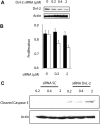
Aberrations of the Wnt canonical pathway (WCP) are known to contribute to the pathogenesis of various types of cancer. We hypothesize that these defects may exist in mantle cell lymphoma (MCL). Both the upstream and downstream aspects of WCP were examined in MCL cell lines and tumors. Using WCP-specific oligonucleotide arrays, we found that MCL highly and consistently expressed Wnt3 and Wnt10. beta-catenin, a transcriptional factor that is a downstream target of WCP, is localized to the nucleus and transcriptionally active in all 3 MCL cell lines examined. By immunohistochemistry, 33 (52%) of 64 MCL tumors showed nuclear localization of beta-catenin, which significantly correlated with the expression of the phosphorylated/inactive form of GSK3beta (p-GSK3beta; P = .011, Fisher). GSK3beta inactivation is directly linked to WCP stimulation, since addition of recombinant sFRP proteins (a naturally occurring decoy for the Wnt receptors) resulted in a significant decrease in p-GSK3beta. Down-regulation of DvL-2 (an upstream signaling protein in WCP) by siRNA or selective inhibition of beta-catenin using quercetin significantly decreased cell growth in MCL cell lines. To conclude, WCP is constitutively activated in a subset of MCL and it appears to promote tumorigenesis in MCL.
Figures


Comment in
-
Wnt-erizing mantle cell lymphoma.Blood. 2008 Dec 15;112(13):4783-4. doi: 10.1182/blood-2008-10-181792. Blood. 2008. PMID: 19064731 No abstract available.
Similar articles
-
Gene Methylation and Silencing of WIF1 Is a Frequent Genetic Abnormality in Mantle Cell Lymphoma.Int J Mol Sci. 2021 Jan 18;22(2):893. doi: 10.3390/ijms22020893. Int J Mol Sci. 2021. PMID: 33477402 Free PMC article.
-
Wnt/β-catenin signaling pathway and thioredoxin-interacting protein (TXNIP) mediate the "glucose sensor" mechanism in metastatic breast cancer-derived cells MDA-MB-231.J Cell Physiol. 2012 Feb;227(2):578-86. doi: 10.1002/jcp.22757. J Cell Physiol. 2012. PMID: 21448924
-
Biological and clinical significance of GSK-3beta in mantle cell lymphoma--an immunohistochemical study.Int J Clin Exp Pathol. 2010 Jan 25;3(3):244-53. Int J Clin Exp Pathol. 2010. PMID: 20224723 Free PMC article.
-
Redundant expression of canonical Wnt ligands in human breast cancer cell lines.Oncol Rep. 2006 Mar;15(3):701-7. Oncol Rep. 2006. PMID: 16465433
-
Distinctive microRNA signature associated of neoplasms with the Wnt/β-catenin signaling pathway.Cell Signal. 2013 Dec;25(12):2805-11. doi: 10.1016/j.cellsig.2013.09.006. Epub 2013 Sep 13. Cell Signal. 2013. PMID: 24041653 Review.
Cited by
-
Increased incidence of endometrioid tumors caused by aberrations in E-cadherin promoter of mismatch repair-deficient mice.Carcinogenesis. 2011 Jul;32(7):1085-92. doi: 10.1093/carcin/bgr080. Epub 2011 May 5. Carcinogenesis. 2011. PMID: 21551128 Free PMC article.
-
Gene Methylation and Silencing of WIF1 Is a Frequent Genetic Abnormality in Mantle Cell Lymphoma.Int J Mol Sci. 2021 Jan 18;22(2):893. doi: 10.3390/ijms22020893. Int J Mol Sci. 2021. PMID: 33477402 Free PMC article.
-
Influence of five potential anticancer drugs on wnt pathway and cell survival in human biliary tract cancer cells.Int J Biol Sci. 2012;8(1):15-29. doi: 10.7150/ijbs.8.15. Epub 2011 Nov 7. Int J Biol Sci. 2012. PMID: 22211101 Free PMC article.
-
Mantle cell lymphoma: biology, pathogenesis, and the molecular basis of treatment in the genomic era.Blood. 2011 Jan 6;117(1):26-38. doi: 10.1182/blood-2010-04-189977. Epub 2010 Oct 12. Blood. 2011. PMID: 20940415 Free PMC article. Review.
-
Stabilization of β-catenin upon B-cell receptor signaling promotes NF-kB target genes transcription in mantle cell lymphoma.Oncogene. 2020 Apr;39(14):2934-2947. doi: 10.1038/s41388-020-1183-x. Epub 2020 Feb 7. Oncogene. 2020. PMID: 32034308
References
-
- Swerdlow SH, F B, Isaacson PI, et al. Mantle cell lymphoma. In: Jaffe ES, Harris NL, Stein H, Vardiman JW, editors. World Health Organization Classification of Tumors: Pathology and Genetics of Tumors of Haematopoetic and Lymphoid Tissues. Lyon, France: IARC Press; 2001. pp. 168–170.
-
- Campo E, Raffeld M, Jaffe ES. Mantle-cell lymphoma. Semin Hematol. 1999;36:115–127. - PubMed
Publication types
MeSH terms
Substances
Grants and funding
LinkOut - more resources
Full Text Sources
Other Literature Sources
Molecular Biology Databases

