

Update on pancreatic intraepithelial neoplasia
- PMID: 18787611
- PMCID: PMC2480542
Update on pancreatic intraepithelial neoplasia
Abstract
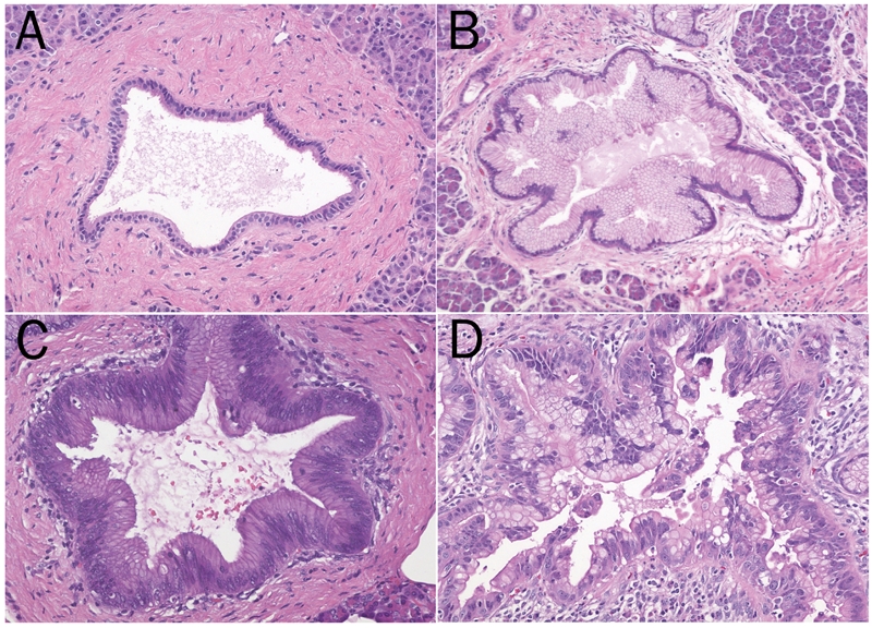
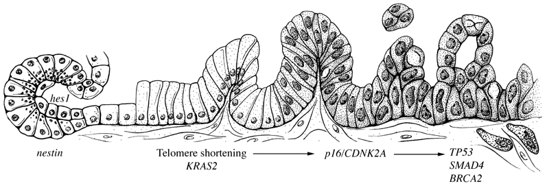
Pancreatic intraepithelial neoplasia (PanIN) is a histologically well-defined precursor to invasive ductal adenocarcinoma of the pancreas. PanINs are remarkably common lesions, particularly in the elderly population. Molecular studies have helped establish the progression of PanIN to invasive cancer, and recently genetically engineered mouse models have been generated that recapitulate the entire spectrum of lesions from precursor to invasive pancreatic cancer. Some PanIN lesions produce lobulocentric atrophy of the pancreatic parenchyma, and, when multifocal, this lobulocentric atrophy may be detectable using currently available imaging techniques such as endoscopic ultrasound. The association of acinar-ductal metaplasia with PanIN lesions has led some to hypothesize that PanINs develop from acinar cells that undergo acinar-ductal metaplasia.
Keywords: Pancreatic cancer; epigenetics; genetics; intraepithelial neoplasm; metaplasia.
Figures
Similar articles
-
Telomeres are shortened in acinar-to-ductal metaplasia lesions associated with pancreatic intraepithelial neoplasia but not in isolated acinar-to-ductal metaplasias.Mod Pathol. 2011 Feb;24(2):256-66. doi: 10.1038/modpathol.2010.181. Epub 2010 Sep 24. Mod Pathol. 2011. PMID: 20871595 Free PMC article.
-
Pancreatic Intraepithelial Neoplasia.Surg Pathol Clin. 2011 Jun;4(2):523-35. doi: 10.1016/j.path.2011.03.005. Epub 2011 May 30. Surg Pathol Clin. 2011. PMID: 26837486 Review.
-
Acinar cells contribute to the molecular heterogeneity of pancreatic intraepithelial neoplasia.Am J Pathol. 2007 Jul;171(1):263-73. doi: 10.2353/ajpath.2007.061176. Am J Pathol. 2007. PMID: 17591971 Free PMC article.
-
Pancreatic intraepithelial neoplasia in the background of invasive ductal carcinoma of the pancreas as a prognostic factor.Histopathology. 2014 Sep;65(3):389-97. doi: 10.1111/his.12397. Epub 2014 Apr 16. Histopathology. 2014. PMID: 24931343
-
Pancreatic intraepithelial neoplasia revisited and updated.Pancreatology. 2009;9(1-2):45-54. doi: 10.1159/000178874. Epub 2008 Dec 12. Pancreatology. 2009. PMID: 19077454 Review.
Cited by
-
Clinical significance of the integrin α6β4 in human malignancies.Lab Invest. 2015 Sep;95(9):976-86. doi: 10.1038/labinvest.2015.82. Epub 2015 Jun 29. Lab Invest. 2015. PMID: 26121317 Free PMC article. Review.
-
A comprehensive review of pancreatic cancer and its therapeutic challenges.Aging (Albany NY). 2022 Sep 28;14(18):7635-7649. doi: 10.18632/aging.204310. Epub 2022 Sep 28. Aging (Albany NY). 2022. PMID: 36173644 Free PMC article. Review.
-
NF-κB and Pancreatic Cancer; Chapter and Verse.Cancers (Basel). 2021 Sep 7;13(18):4510. doi: 10.3390/cancers13184510. Cancers (Basel). 2021. PMID: 34572737 Free PMC article. Review.
-
Tumor Dormancy and Relapse: From a Natural Byproduct of Evolution to a Disease State.Cancer Res. 2017 May 15;77(10):2564-2569. doi: 10.1158/0008-5472.CAN-17-0068. Cancer Res. 2017. PMID: 28507050 Free PMC article. Review.
-
Molecular signatures of pancreatic cancer.Arch Pathol Lab Med. 2011 Jun;135(6):716-27. doi: 10.5858/2010-0566-RA.1. Arch Pathol Lab Med. 2011. PMID: 21631264 Free PMC article. Review.
References
-
- American Cancer Society. Cancer. New York, New York: American Cancer Society; 2007. Cancer Facts and Figures 2007; pp. 1–52.
-
- Berry DA, Cronin KA, Plevritis SK, Fryback DG, Clarke L, Zelen M, Mandelblatt JS, Yakovlev AY, Habbema JD, Feuer EJ. Effect of screening and adjuvant therapy on mortality from breast cancer. N Engl J Med. 2005;353:1784–1792. - PubMed
-
- Faivre J, Dancourt V, Lejeune C, Tazi MA, Lamour J, Gerard D, Dassonville F, Bonithon-Kopp C. Reduction in colorectal cancer mortality by fecal occult blood screening in a French controlled study. Gastroenterology. 2004;126:1674–1680. - PubMed
-
- Frazier AL, Colditz GA, Fuchs CS, Kuntz KM. Cost-effectiveness of screening for colorectal cancer in the general population. JAMA. 2000;284:1954–1961. - PubMed
-
- Newcomb PA, Storer BE, Morimoto LM, Templeton A, Potter JD. Long-term efficacy of sigmoidoscopy in the reduction of colorectal cancer incidence. J Natl Cancer Inst. 2003;95:622–625. - PubMed
LinkOut - more resources
Full Text Sources
Molecular Biology Databases