. 2008 Jan 1;1(5):387-95.

Hematopoietic disorders in Down syndrome

Affiliations

Hematopoietic disorders in Down syndrome

John K Choi. Int J Clin Exp Pathol. .

Abstract

Patients with Down syndrome have an increased risk of developing various hematological disorders. In this article, the clinical characteristics and differential diagnosis of the hematological disorders associated with Down syndrome are reviewed, and the underlying molecular mechanisms discussed.

Keywords: Down syndrome; acute leukemia; myelodysplastic syndrome; transient myeloproliferative disorder.


Figures
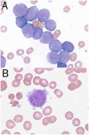
Figure 1
Figure 1
Cytology of the megakaryoblasts in TMD and AMKL (1000× magnification). A. Large sized megakaryoblasts with irregular nuclei and moderate pale blue cytoplasm resembling myeloblasts. B. Typical megakaryoblastic morphology with cytoplasmic projections and tiny azurophilic platelet granules.
Figure 2
Figure 2
Usual cytochemical staining patterns of megakaryoblasts in TMD and AMKL (1000× magnification). A. Sudan Black B. Similar negative staining is seen with myeloperoxidase and chloroacetate esterase. B. Acid phosphatase. C. Non-specific esterase using conditions resulting in diffuse cytoplasmic staining of monocytes and macrophages. Megakaryoblast (upper left) is negative while macrophage (lower right) is strongly and diffusely positive. D. Non-specific esterase using conditions resulting in multi-punctate staining of megakaryoblasts. This pattern is partially resistant to sodium fluoride.
Figure 3
Figure 3
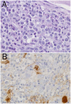
Bone marrow biopsy findings in AMKL. A. Extensive infiltration of the marrow by AMKL blasts (PAS stain, 400× magnification). B. Immunohistochemistry stain for Factor VIII-related antigen is negative in the AMKL blasts despite strong reactivities seen in megakaryocytes (400× magnification).
Figure 4
Figure 4
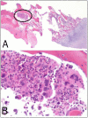
Focal megakaryocytic hyperplasia with megakaryocytic dysplasia (paratrabecular localization, clustering, and micromegakaryocytic forms) in a 6 month old DS patient (H&E, 20× (A) and 200 × (B) magnifications, respectively). Circled region in A contains the focal megakaryocytic hyperplasia represented in B. Patient had unexplained cytopenia without increased blasts but developed AMKL one month later.

References

    1. Centers for Disease Control and Prevention. Improved national prevalence estimates for 18 selected major birth defects–United States, 1999–2001. MMWR Morb Mortal Wkly Rep. 2006;54:1301–1305. - PubMed
    1. Henry E, Walker D, Wiedmeier SE, Christensen RD. Hematological abnormalities during the first week of life among neonates with Down syndrome: Data from a multihospital healthcare system. Am J Med Genet A. 2007;143:42–50. - PubMed
    1. Hook EB, Cross PK, Schreinemachers DM. Chromosomal abnormality rates at amniocentesis and in live-born infants. JAMA. 1983;249:2034–2038. - PubMed
    1. Oudejans CB, Go AT, Visser A, Mulders MA, Westerman BA, Blankenstein MA, van Vugt JM. Detection of chromosome 21-encoded mRNA of placental origin in maternal plasma. Clin Chem. 2003;49:1445–1449. - PubMed
    1. Wataganara T, Bianchi DW. Fetal cell-free nucleic acids in the maternal circulation: new clinical applications. Ann N Y Acad Sci. 2004;1022:90–99. - PubMed

LinkOut - more resources