Giant cell tumor of bone: a neoplasm or a reactive condition?
- PMID: 18787633
- PMCID: PMC2480584
Giant cell tumor of bone: a neoplasm or a reactive condition?
Abstract
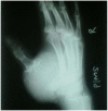
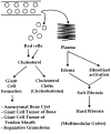
Giant cell tumor of bone (GCTB) is a benign but locally aggressive bone tumor of young adults. It typically presents as a large lytic mass at the end of the epiphysis of long bones. Grossly it is comprised of cystic and hemorrhagic areas with little or no periosteal reaction. Microscopically areas of frank hemorrhage, numerous multinucleated giant cells and spindly stromal cells are present. Telomeric fusions, increased telomerase activity and karyotypic aberrations have been advanced as a proof of its neoplastic nature. However such findings are not universal and can be seen in rapidly proliferating normal cells as well as in several osseous lesions of developmental and/or reactive nature, and the true neoplastic nature of GCTB remains controversial. The ancillary studies have generally not reached to the point where these alone can be taken as sole diagnostic and discriminatory criteria. While giant cells and stromal cells have been extensively studied, little attention has been paid to the overwhelming hemorrhagic component. If examined carefully intact and partially degenerated red blood cells are almost invariably seen in many giant cells as well as in the stroma. While hemorrhage in many patients may be resolved without leaving any trace over time, in some it gives rise to giant cell formation, and in others it may lead to proliferation of fibroblasts and histiocytes. At times one sees xanthomatous cells due to intracytoplasmic cholesterol deposits and sharp cholesterol clefts. Individual genetic makeup, local tissue factors as well as the amount of hemorrhage may play a key role in the final effects and outcome. Malignancy usually does not occur in GCTB and when discover, it usually represents primary bone sarcomas missed at original diagnosis. Embolization therapy to curtail hemorrhage and insertion of cement substance to support matrix are helpful in reducing recurrences. Aneurysmal bone cyst (ABC) shares many features with GCTB. There had been unique karyotypic changes in some aneurysmal bone cysts making it distinct from GCTB. However these changes may be in the endothelial cells which are quite different from stromal or giant cells. It had been concluded that the poor matrix support to the vessels may lead to frequent and profuse intraosseous hemorrhage attracting blood-derived monocytes with active conversion into osteoclasts, resulting in GCTB formation. On the other hand, dilatation of the thin-walled blood vessels results in formation of ABCs. If hemorrhagic foci are replaced by proliferation of fibroblasts and histiocytes, then a picture of fibrous histiocytic lesion is emerged. Enhanced telomerase activity and karyotypic aberrations may be necessary for rapid division of the nuclei of the giant cells in order to be able to deal with significant in situ intraosseous hemorrhage.
Keywords: Giant cell tumor; aneurysmal bone cyst; bone; bone matrix; hemorrhage; osteoclast; osteoclastoma; telomerase.
Figures


Similar articles
-
Prognostic impact of reduced connexin43 expression and gap junction coupling of neoplastic stromal cells in giant cell tumor of bone.PLoS One. 2015 May 1;10(5):e0125316. doi: 10.1371/journal.pone.0125316. eCollection 2015. PLoS One. 2015. PMID: 25933380 Free PMC article.
-
Significance of Biogenetic Markers in Giant Cell Tumor Differentiation and Prognosis: A Narrative Review.Diagnostics (Basel). 2024 Dec 27;15(1):39. doi: 10.3390/diagnostics15010039. Diagnostics (Basel). 2024. PMID: 39795567 Free PMC article. Review.
-
Stromal cells of giant cell tumor of bone show primary cilia in giant cell tumor of bone.Microsc Res Tech. 2022 Mar;85(3):1065-1074. doi: 10.1002/jemt.23976. Epub 2021 Nov 10. Microsc Res Tech. 2022. PMID: 34761465
-
Spectrum of histological features of Denosumab treated Giant Cell Tumor of Bone; potential pitfalls and diagnostic challenges for pathologists.Ann Diagn Pathol. 2020 Apr;45:151479. doi: 10.1016/j.anndiagpath.2020.151479. Epub 2020 Feb 14. Ann Diagn Pathol. 2020. PMID: 32088577
-
Giant-cell-poor giant cell tumor of bone: report of two cases and literature review.Skeletal Radiol. 2023 Sep;52(9):1791-1798. doi: 10.1007/s00256-023-04292-w. Epub 2023 Feb 14. Skeletal Radiol. 2023. PMID: 36781420 Free PMC article. Review.
Cited by
-
Giant cell tumor of the mandible.J Oral Maxillofac Pathol. 2015 Jan-Apr;19(1):108. doi: 10.4103/0973-029X.157217. J Oral Maxillofac Pathol. 2015. PMID: 26097323 Free PMC article.
-
Giant cell tumor of bone and secondary osteoarthritis.Heliyon. 2024 May 9;10(10):e30890. doi: 10.1016/j.heliyon.2024.e30890. eCollection 2024 May 30. Heliyon. 2024. PMID: 38807896 Free PMC article. Review.
-
Rare Presentation of Giant Cell Tumor in the Internal Auditory Canal: Case Report and Review of the Literature.J Neurol Surg Rep. 2015 Jul;76(1):e65-71. doi: 10.1055/s-0034-1396656. Epub 2015 Feb 18. J Neurol Surg Rep. 2015. PMID: 26251814 Free PMC article.
-
Giant cell tumour of bone: new treatments in development.Clin Transl Oncol. 2015 Jun;17(6):419-30. doi: 10.1007/s12094-014-1268-5. Epub 2015 Jan 24. Clin Transl Oncol. 2015. PMID: 25617146 Free PMC article. Review.
-
The Great Need of a Biomechanical-Based Approach for Surgical Methods of Giant Cell Tumor: A Critical Review.J Med Biol Eng. 2017;37(4):454-467. doi: 10.1007/s40846-017-0278-5. Epub 2017 Jun 22. J Med Biol Eng. 2017. PMID: 29541012 Free PMC article. Review.
References
-
- Zheng MH, Robbins P, Xu J, Huang L, Wood DJ, Papadimitriou JM. The histogenesis of giant cell tumour of bone: a model of interaction between neoplastic cells and osteoclasts. Histol Histopathol. 2001;16:297–307. - PubMed
-
- Giant Cell Tumor of Bone. 2004. Available at: http://orthoinfo.aaos.org/fact/thr_report.cfm?Thread_ID=475&topcategory=.Accessed July 20, 2007.
-
- Cheng JC, Johnston JO. Giant cell tumor of bone. prognosis and treatment of pulmonary metastases. Clin Orthop Relat Res. 1997;338:205–214. - PubMed
-
- McGough RL, Rutledge J, Lewis VO, Lin PP, Yasko AW. Impact severity of local recurrence in giant cell tumor of bone. Clin Orthop Relat Res. 2005;438:116–122. - PubMed
LinkOut - more resources
Full Text Sources
