. 2008 Sep 12;4(9):e1000144.
doi: 10.1371/journal.ppat.1000144.

Listeriolysin S, a novel peptide haemolysin associated with a subset of lineage I Listeria monocytogenes

Affiliations

Listeriolysin S, a novel peptide haemolysin associated with a subset of lineage I Listeria monocytogenes

Paul D Cotter et al. PLoS Pathog. .

Abstract

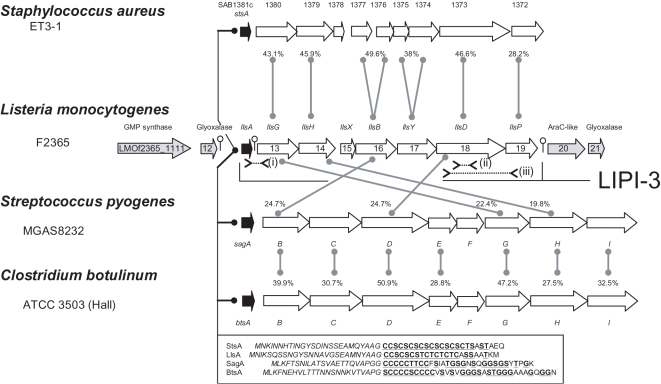
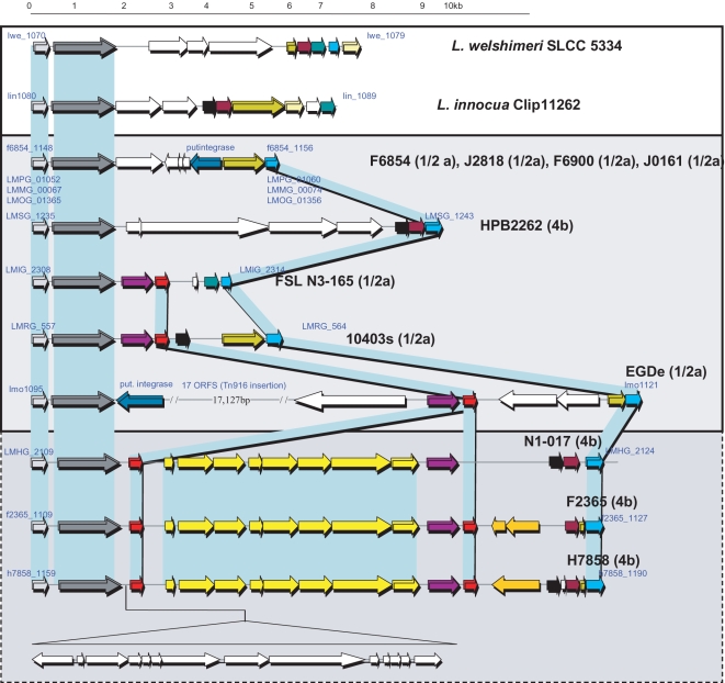
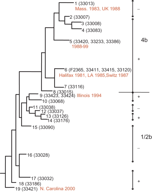
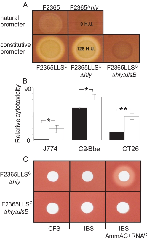
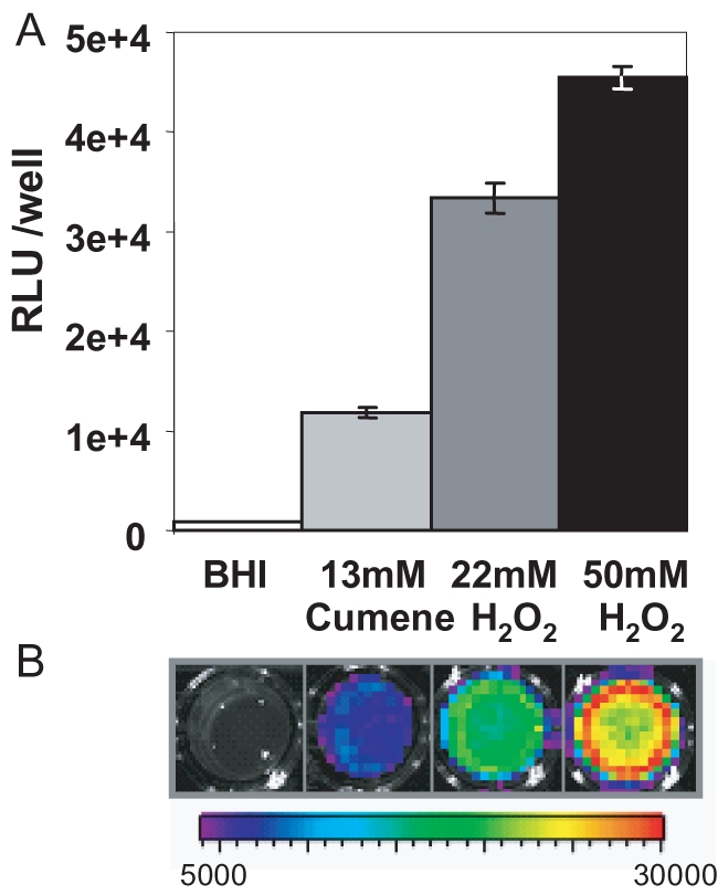
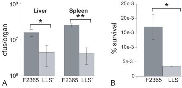
Streptolysin S (SLS) is a bacteriocin-like haemolytic and cytotoxic virulence factor that plays a key role in the virulence of Group A Streptococcus (GAS), the causative agent of pharyngitis, impetigo, necrotizing fasciitis and streptococcal toxic shock syndrome. Although it has long been thought that SLS and related peptides are produced by GAS and related streptococci only, there is evidence to suggest that a number of the most notorious Gram-positive pathogenic bacteria, including Listeria monocytogenes, Clostridium botulinum and Staphylococcus aureus, produce related peptides. The distribution of the L. monocytogenes cluster is particularly noteworthy in that it is found exclusively among a subset of lineage I strains; i.e., those responsible for the majority of outbreaks of listeriosis. Expression of these genes results in the production of a haemolytic and cytotoxic factor, designated Listeriolysin S, which contributes to virulence of the pathogen as assessed by murine- and human polymorphonuclear neutrophil-based studies. Thus, in the process of establishing the existence of an extended family of SLS-like modified virulence peptides (MVPs), the genetic basis for the enhanced virulence of a proportion of lineage I L. monocytogenes may have been revealed.


Conflict of interest statement

The authors have declared that no competing interests exist.

Figures

Figure 1
Figure 1. Comparison of MVP-associated operons.
Arrangement of the Stapholysin S (sts), Listeriolysin S (lls), Streptolysin S (sag) and Botulysin (bts) associated genes in S. aureus ET3-1, L. monocytogenes F2365, S. pyogenes MGAS 8232 and C. botulinum ATCC3502 respectively. Homology between the predicted lls gene products and their sag and sts equivalents is indicated by a line and dashed line, respectively, with an associated % identity value. % identity values with respect to the predicted Sag and Bts gene products are also indicated. The amino acid sequence of the predicted unmodified structural peptides, StsA, LlsA, SagA and BtsA is presented (boxed) with italicized letters indicating amino acids likely to constitute the leader region. Residues within the structural propeptide that are potentially modified are underlined. The location of oligonucleotide pairs used to investigate strain variation are indicated (i) llsAint-llsAsoeD, (ii) 1118degf–1118degr and (iii) 1118f–1118r.
Figure 2
Figure 2. LIPI-3 and corresponding regions in LIPI-3 minus Listeria.
Comparison of the LIPI-3-containing region (bottom) with the corresponding region of LLS L. monocytogenes (middle), L. innocua and L. welshimeri . For L. monocytognes strains, the strain name and serotype (in brackets) is presented and the designation of the first and last gene in each case is that designated in the corresponding genome sequence. Homologous genes (or in the case of the llsA-P, clusters of genes) are presented by matching colours.
Figure 3
Figure 3. Distribution of LIPI-3 among lineage I L. monocytogenes.
LIPI-3 status (positive, + or negative, −) mapped onto a maximum parsimony tree inferred from sequence data of lineage I strains (adapted from [24]). The individual sequence types (1–19), representatives tested (strain number in brackets) and associated epidemic outbreaks (in red). Strains of serotype 4b and 1/2b are also distinguished.
Figure 4
Figure 4. Haemolytic and cytolytic activities of LLS.
(A) Haemolysis of Columbia blood agar (5% Sheep's blood) by F2365, F2365Δhly and F2365ΔhlyΔllsB when the lls genes is under the control of the natural PllsA or constitutive PHELP promoters (F2365LLSC, F2365LlsCΔhly and F2365LLSCΔhlyΔllsB). In situations where haemolytic activity was quantified, the corresponding haemolytic unit (H.U.) values are presented. (B) Cytotoxicity relative to F2365 (100%) with respect to the J774, C2-Bbe and CT26 cell lines; F2365Δhly-black, F2365LLSCΔhly-white. *-significantly different (P<0.05), **-extremely significantly different (P<0.005) different. Error bars represent standard error of the mean. (C) Assessment of the haemolytic activity of cell free supernatant (CFS) of F2365LLSCΔhly and F2365LLSCΔhlyΔllsB, of the cell free supernatant of induction buffer washed F2365LLSCΔhly and F2365LLSCΔhlyΔllsB (IBS), and of IBS from the strains combined with RNA core (RNAC; inducer) and ammonium acetate (AmmAc; stabilizer) aliquoted into 4.6 mm wells in Columbia blood agar (5% Sheep's blood). NB. IB, RNAC and AmmAc, both individually and in combination, are non-haemolytic (data not shown).
Figure 5
Figure 5. Induction of the PllsA promoter.
Induction of PllsA by 13 mM cumene hydroperoxide and 22 and 50 mM hydrogen peroxide. Data are presented as (A) mean relative light units (RLU; photons s−1)/well±standard deviations for three replicates, and (B) one representative of three independent experiments is shown. The colour bar indicates bioluminescence signal intensity (in photons s−1 cm−2).
Figure 6
Figure 6. Comparison of the virulence of wildtype F2365 and a LLS derivative.
(A) Levels (cfu–colony forming units) of wild-type (F2365) and mutant (F2365ΔllsB; LLS) F2365 in livers and spleens of Balb/C mice 3 days post-intraperitoneal infection. (B) Survival in human PMNs after 2 hr. * - significantly different (P<0.05), ** - extremely significantly different (P<0.005) different. Error bars represent standard error of the mean.

References

    1. Xie L, van der Donk WA. Post-translational modifications during lantibiotic biosynthesis. Curr Opin Chem Biol. 2004;8:498–507. - PubMed
    1. Jack RW, Jung G. Lantibiotics and microcins: polypeptides with unusual chemical diversity. Curr Opin Chem Biol. 2000;4:310–317. - PubMed
    1. Cotter PD, Hill C, Ross RP. Food Microbiology: Bacteriocins: developing innate immunity for food. Nat Rev Microbiol. 2005;3:777–788. - PubMed
    1. Cox CR, Coburn PS, Gilmore MS. Enterococcal Cytolysin: A Novel Two Component Peptide System that Serves as a Bacterial Defense Against Eukaryotic and Prokaryotic Cells. Curr Protein Pept Sci. 2005;6:77–84. - PubMed
    1. Datta V, Myskowski SM, Kwinn LA, Chiem DN, Varki N, et al. Mutational analysis of the group A streptococcal operon encoding streptolysin S and its virulence role in invasive infection. Mol Microbiol. 2005;56:681–695. - PubMed

Publication types

Substances