. 2008 Sep 12;4(9):e1000191.
doi: 10.1371/journal.pgen.1000191.

Evolution of a new function by degenerative mutation in cephalochordate steroid receptors

Affiliations

Evolution of a new function by degenerative mutation in cephalochordate steroid receptors

Jamie T Bridgham et al. PLoS Genet. .

Abstract

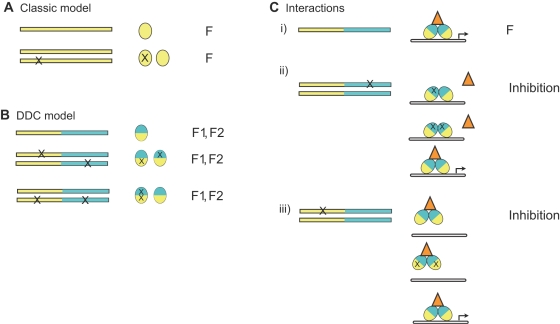
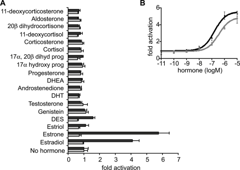
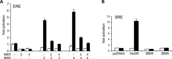
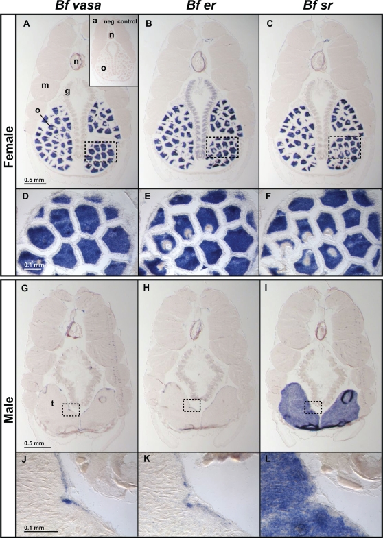
Gene duplication is the predominant mechanism for the evolution of new genes. Major existing models of this process assume that duplicate genes are redundant; degenerative mutations in one copy can therefore accumulate close to neutrally, usually leading to loss from the genome. When gene products dimerize or interact with other molecules for their functions, however, degenerative mutations in one copy may produce repressor alleles that inhibit the function of the other and are therefore exposed to selection. Here, we describe the evolution of a duplicate repressor by simple degenerative mutations in the steroid hormone receptors (SRs), a biologically crucial vertebrate gene family. We isolated and characterized the SRs of the cephalochordate Branchiostoma floridae, which diverged from other chordates just after duplication of the ancestral SR. The B. floridae genome contains two SRs: BfER, an ortholog of the vertebrate estrogen receptors, and BfSR, an ortholog of the vertebrate receptors for androgens, progestins, and corticosteroids. BfSR is specifically activated by estrogens and recognizes estrogen response elements (EREs) in DNA; BfER does not activate transcription in response to steroid hormones but binds EREs, where it competitively represses BfSR. The two genes are partially coexpressed, particularly in ovary and testis, suggesting an ancient role in germ cell development. These results corroborate previous findings that the ancestral steroid receptor was estrogen-sensitive and indicate that, after duplication, BfSR retained the ancestral function, while BfER evolved the capacity to negatively regulate BfSR. Either of two historical mutations that occurred during BfER evolution is sufficient to generate a competitive repressor. Our findings suggest that after duplication of genes whose functions depend on specific molecular interactions, high-probability degenerative mutations can yield novel functions, which are then exposed to positive or negative selection; in either case, the probability of neofunctionalization relative to gene loss is increased compared to existing models.


Conflict of interest statement

The authors have declared that no competing interests exist.

Figures

Figure 1
Figure 1. Effect of degenerative mutations on the function of a duplicate gene that depends on molecular interactions.
A) In the classic model, duplicate genes are redundant and do not interact, so mutation in one copy causes no change in the function (F) and evolve neutrally. B) In the DDC model, duplicate genes, which have two or more modular subfunctions (F1 and F2, blue and yellow), are redundant and do not interact. Degenerative mutations in subfunctions cause no change in function if the subfunction is retained in the other copy and evolve neutrally. C) If a gene depends upon molecular interactions for its function, degenerative mutations in one copy can affect the function of the other and therefore be selected for or against. i) In the case shown, a gene product must dimerize and interact with DNA (white bar) and an accessory factor (orange triangle) for its function (transcriptional activation). ii) After duplication, degenerative mutations that impair the interactions of one copy with the accessory factor yield a nonfunctional product that competes for DNA binding sites, reducing the activity of the other copy. iii) Degenerative mutations that impair DNA binding in one copy yield nonfunctional products that compete for accessory factors and reduce the activity of the other copy. Inhibition also occurs if the duplicate genes do not dimerize but compete for other binding partners.
Figure 2
Figure 2. Cephalochordates have one ortholog each of the ER and kSR subfamilies.
A) Sequence similarity of BfER and BfSR to each other and human steroid receptors. The percent of identical amino acid residues for each pairwise comparison is shown. DBD and LBD, DNA-binding and ligand-binding domains. B) Reduced phylogeny of the steroid receptor gene family. Numbers in parentheses refer to the number of sequences in each group used in the analysis. Estrogen-sensitive receptors, including the ancestral steroid receptor (AncSR1) are in blue; ketosteroid receptors are in yellow. Black triangle, loss of transcriptional activation; yellow triangle, gain of ketosteroid sensitivity in the vertebrate AR/PR/GR/MR clade. Support for each branch is shown as the approximate likelihood ratio (the ratio of the likelihood of the best tree with that node to the best tree without it), the chi-square likelihood confidence statistic for the node, and the Bayesian posterior probability. Nodes that place B. floridae receptors as orthologs of the ERs and kSRs are in red. Scale bar shows expected per-site substitution rate for branch lengths. Complete phylogenies and a list of genes, species, and accessions are in Figures S6, S7, S8 and Table S1. C) Conserved synteny between BfER-containing scaffold 42 and human chromosomes 14 and 6, which contain ERβ (ESR2) and ERα (ESR1), respectively. Green boxes with connecting lines indicate reciprocal BLAST best-hits between scaffold 42 in the B. floridae genome and human chromosome 14; purple boxes, orthologs between Bf scaffold 42 and human chromosome 6. Genes are shown in order with spacing approximately proportional to physical distance. BfER, ESR1, and ESR2 all share orthologous nearest neighbors (red lines).
Figure 3
Figure 3. Estrogens activate BfSR but not BfER.
A) Hormone-activated transcription of a luciferase reporter gene by the ligand-binding domains of BfSR (black) and BfER (gray) is shown as fold activation relative to vehicle-only control. All hormone treatments were at 1 µM concentration. DES, diethylstilbesterol; DHT, dihydrotestosterone; DHEA, dehydroepiandrosterone; Prog, progesterone. Mean+/−SE of three replicates is shown. B) Dose-response relationship for transcriptional activation by BfSR LBD in the presence of increasing concentrations of estradiol (black) and estrone (gray).
Figure 4
Figure 4. BfER represses BfSR-mediated transcription on estrogen response elements.
A) Full-length BfSR activates transcription of an ERE-driven reporter gene in the presence of 1 µM estradiol (black bars), but activation is inhibited by transfection of increasing quantities of BfER. The quantity of each plasmid transfected is indicated in ng/well. White bars, vehicle-only (no hormone) control. B) Neither BfER nor BfSR activates reporter transcription from an SRE binding site. Cells were transfected with an SRE-driven luciferase reporter and full-length BfER, BfSR, empty vector (pcDNA3), or positive control (HsGR; human GR). Black bars, hormone treatment at 1 uM; white bars, vehicle only.
Figure 5
Figure 5. BfER repression of BfSR by competition for EREs.
A) BfER's DNA-binding domain recognizes an estrogen response element (ERE). A fusion of the BfER-DBD with a constitutive activation domain activates transcription of an ERE-driven luciferase reporter gene. Negative control (pCMV-AD vector only) and positive control (HsER, human ERα DBD) are shown for comparison. Replacement C205A in the BfER-DBD abolishes recognition of an ERE. B) Neither the BfER-DBD nor the BfSR-DBD recognize steroid response elements (SRE). Negative control (pCMV-AD vector only) and positive control (HsGR, human GR DBD) are shown for comparison. C) BfER and BfSR bind directly to EREs. An electromobility shift assay shows that each receptor binds to a biotin-labeled ERE and migrates as expected (BfER, 521 amino acids, ∼58 kD; BfSR, 616 amino acids, ∼68 kD). When the two receptors are co-transfected, ERE-binding by the BfER homodimer is so strong that it is unclear whether BfER and BfSR heterodimerize. D) Mutation C205A, which compromises DNA binding, abrogates BfER's capacity to repress BfSR-mediated transcription of an ERE-driven reporter. Full-length BfSR, wild-type BfER (BfER-WT), and the BfER C205A mutant were cotransfected. Cells were treated with no hormone (white bars) or 1 µM estradiol (black bars).
Figure 6
Figure 6. BfER and BfSR are co-expressed.
Alternating cross-sections of adult animals were probed with digoxigenin-labelled RNAs for Bfer, Bfsr, or Vasa, a germ cell marker. A–F) In adult females, expression of Bfer and Bfsr are co-localized in the cytoplasm of oocytes in the ovary (o); er is also weakly expressed in the female gill bars (g). The negative control experiment with no probe (inset in A) shows no endogenous alkaline phosphatase activity. G–L) In adult males Bfer and Vasa are expressed only in a single cell layer of early spermatogonia in the testicular epithelium, but Bfsr is expressed throughout testes (t). Myotome (m) and notochord (n) are also labeled. Scale bars are indicated.
Figure 7
Figure 7. Mechanism for evolution of a duplicate receptor repressor.
A) Conservation and variability of ligand-contacting residues. The figure shows a generic steroid hormone surrounded by the amino acids that line the ligand-binding pocket in the crystal structures of the human ERα. For each site, residues from the human ERα (HsER), BfSR, BfER, and AncSR1 are shown, from top to bottom, numbered according to the human ERα sequence. Moieties at the 3 and 17 positions, shown as large circles, vary among steroids. B) Historical substitutions R394C and F404L, which occurred in the lineage leading to transcriptional repressor BfER, are predicted to abolish estrogen binding and activation. Left, x-ray crystal structure of the ligand pocket of the human ERα with estradiol. Arg394 and Phe404 play key roles in a network of hydrogen bonds and packing interactions that stabilize the ligand and transcriptionally active conformation. Right, mutations R394C and F404L from BfER disrupt this network. Red sphere, water molecule. C) Mutations R394C and F404L, introduced into BfSR, abolish the receptor's transcriptional capacity and generate a dose-dependent repressor of the wild-type (WT) BfSR. Numbers show the quantity of each plasmid transfected (in ng) with 1 µM estradiol (black bars) or with no hormone added (vehicle only, white bars). Mutation C205A, which disrupts DNA-binding, eliminates BfER's capacity to repress BfSR-driven transcription. D) Either mutation R394C or F404L, introduced singly into BfSR, are each sufficient to abolish transcriptional capacity and generate a repressor of BfSR-WT.

References

    1. Orengo CA, Thornton JM. Protein families and their evolution-a structural perspective. Annu Rev Biochem. 2005;74:867–900. - PubMed
    1. Muller A, MacCallum RM, Sternberg MJ. Structural characterization of the human proteome. Genome Res. 2002;12:1625–1641. - PMC - PubMed
    1. Teichmann SA, Babu MM. Gene regulatory network growth by duplication. Nat Genet. 2004;36:492–496. - PubMed
    1. Presser A, Elowitz MB, Kellis M, Kishony R. The evolutionary dynamics of the Saccharomyces cerevisiae protein interaction network after duplication. Proc Natl Acad Sci U S A. 2008;105:950–954. - PMC - PubMed
    1. Haldane JBS. The part played by recurrent mutation in evolution. American Naturalist. 1933;67:5–19.

Publication types