

Effect of blood brain barrier permeability in recurrent high grade gliomas on the intratumoral pharmacokinetics of methotrexate: a microdialysis study
- PMID: 18787762
- PMCID: PMC4029102
- DOI: 10.1007/s11060-008-9678-2
Effect of blood brain barrier permeability in recurrent high grade gliomas on the intratumoral pharmacokinetics of methotrexate: a microdialysis study
Abstract
Purpose: Determining whether potentially therapeutic drug exposure is achieved within brain tumors in an exploratory clinical investigation would provide a rational basis for selecting agents for evaluation in phase II trials. This study investigated the use of microdialysis to assess intratumoral drug distribution in patients with recurrent high grade gliomas (HGG).
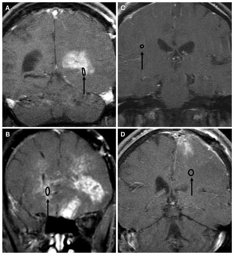
Patients and methods: Microdialysis catheters were placed during surgery for residual HGG 1-day before giving methotrexate (MTX) 12-g/m(2) by 4-h i.v. infusion. MTX was measured by Liquid Chromatography/Mass Spectrometry (LC/MS) in plasma and microdialysate during the infusion and for 24-h thereafter. Blood brain barrier (BBB) permeability of tissue in which the microdialysis probe was located was determined by digitally fusing brain CT and contrast enhanced MRI images.
Results: The microdialysis probe was located in contrast enhancing tumor in two patients and nonenhancing tissue in two others. Cerebral drug penetration, as indicated by the ratio of the area under the MTX concentration-time curves in brain extracellular fluid and plasma, was considerably greater in contrast enhancing tumor (0.28-0.31) than nonenhancing tissue (0.032-0.094). Nevertheless, MTX concentrations in ECF exceeded 2-microM, the average concentration for 50% cell kill against glioma cell lines in vitro, for 20-26 h in both regions of the tumor.
Conclusions: Microdialysis is a very informative technique for characterizing the intratumoral pharmacokinetics of drugs, such as MTX, that do not freely penetrate the BBB. Establishing the catheter probe location relative to areas of BBB disruption is required to properly assess the significance of microdialysis data in this context.
Figures

References
-
- Brandsma D, van den Bent MJ. Molecular targeted therapies and chemotherapy in malignant gliomas. Curr Opin Oncol. 2007;19(6):598–605. - PubMed
Publication types
MeSH terms
Substances
Grants and funding
LinkOut - more resources
Full Text Sources
Medical
