. 2008 Nov 1;323(1):6-18.
doi: 10.1016/j.ydbio.2008.08.019. Epub 2008 Aug 29.

Development of the vertebral morphogenetic field in the mouse: interactions between Crossveinless-2 and Twisted Gastrulation

Affiliations

Development of the vertebral morphogenetic field in the mouse: interactions between Crossveinless-2 and Twisted Gastrulation

Lise Zakin et al. Dev Biol. .

Abstract

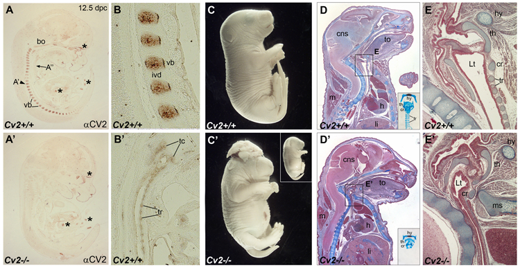
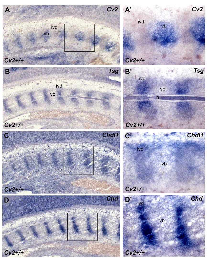
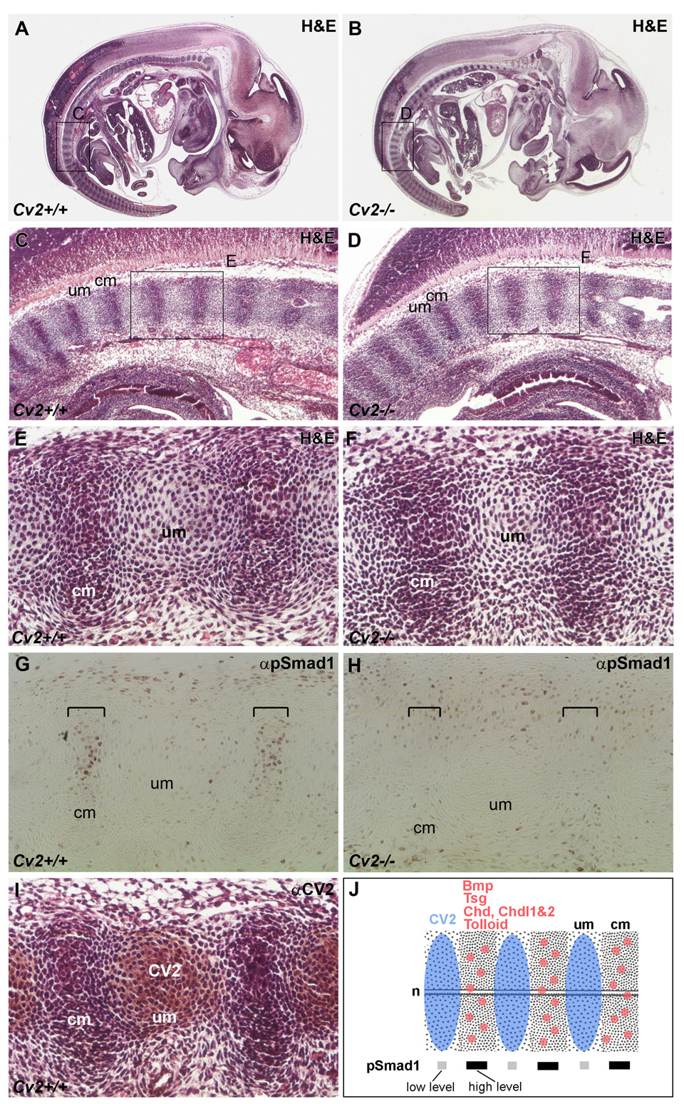
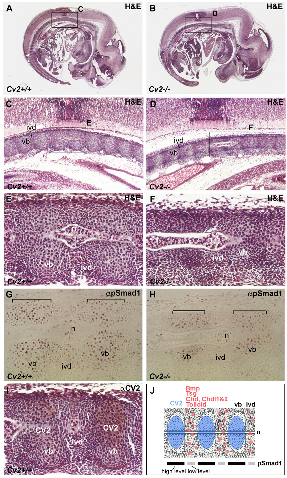
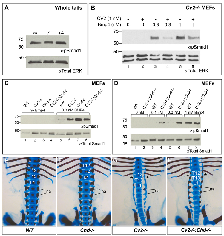
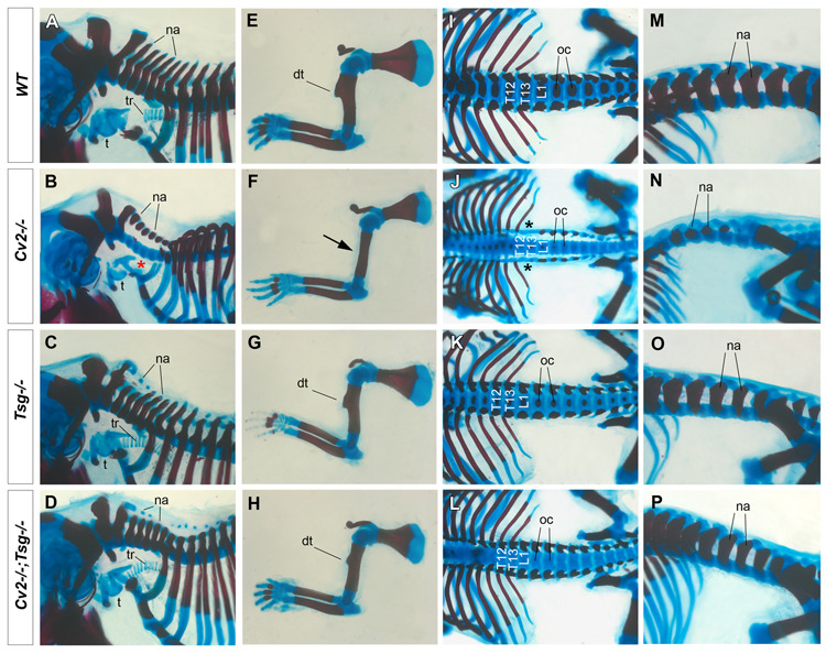
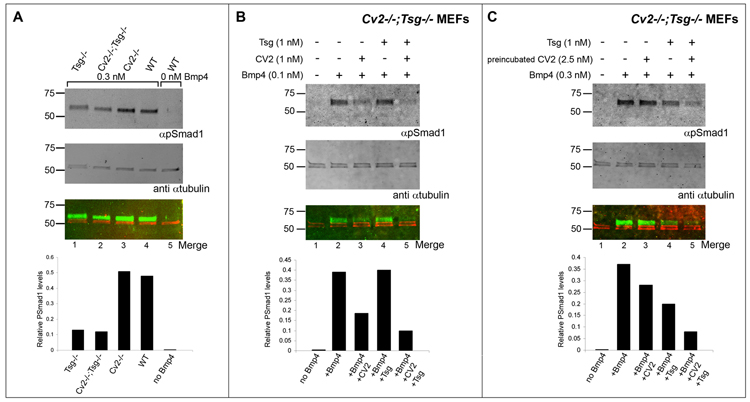
Crossveinless-2 (Cv2), Twisted Gastrulation (Tsg) and Chordin (Chd) are components of an extracellular biochemical pathway that regulates Bone Morphogenetic Protein (BMP) activity during dorso-ventral patterning of Drosophila and Xenopus embryos, the formation of the fly wing, and mouse skeletogenesis. Because the nature of their genetic interactions remained untested in the mouse, we generated a null allele for Cv2 which was crossed to Tsg and Chd mutants to obtain Cv2; Tsg and Cv2; Chd compound mutants. We found that Cv2 is essential for skeletogenesis as its mutation caused the loss of multiple bone structures and posterior homeotic transformation of the last thoracic vertebra. During early vertebral development, Smad1 phosphorylation in the intervertebral region was decreased in the Cv2 mutant, even though CV2 protein is normally located in the future vertebral bodies. Because Cv2 mutation affects BMP signaling at a distance, this suggested that CV2 is involved in the localization of the BMP morphogenetic signal. Cv2 and Chd mutations did not interact significantly. However, mutation of Tsg was epistatic to all CV2 phenotypes. We propose a model in which CV2 and Tsg participate in the generation of a BMP signaling morphogenetic field during vertebral formation in which CV2 serves to concentrate diffusible Tsg/BMP4 complexes in the vertebral body cartilage.


Figures

Fig. 1
Fig. 1
CV2 expression pattern at 12.5 d.p.c. and Cv2−/− phenotype. (A-A’) Detection of the CV2 protein on sagittal paraffin sections of wild-type and Cv2 mutant mouse embryos at 12.5 d.p.c. by immunohistochemistry. Note in wild-type the CV2 protein staining in the vertebral bodies (vb). No CV2 protein is detected in Cv2−/− embryos. Asterisks indicate unspecific signals present in both wild-type and mutant embryos. bo, basioccipital bone. (B) Higher magnification view of the CV2 staining in the vertebral bodies. ivd, intervertebral disc. (B’) The CV2 signal in thyroid cartilages (tc) and tracheal rings (tr), shown at a higher magnification. (C-C’) External view of littermates from Cv2+/− intercrosses showing shorter tails in Cv2−/− compared to wild-type and exencephaly observed in 25% of the mutants. Inset shows a Cv2−/− mutant lacking exencephaly. (D-E’) Mallory’s tetrachrome staining of sagittal paraffin sections of wild-type (top panels) and mutant (bottom panels) neonates. Cv2−/− embryos die at birth of respiratory failure. Note in the thoracic region of Cv2−/− the reduction of the lumen of the trachea and the decreased distance between the vertebral column and the manubrium of the sternum (ms). Insets show tracheas of wild-type and Cv2−/− embryos stained with Alcian Blue and Alizarin Red. Note that in the mutant the cartilage rings that support the trachea are absent. Boxed areas are shown at higher magnification in (E) and (E’). In the mutant the collapse of the tracheal lumen (Lt) is due to the absence of the tracheal rings (tr). cns, central nervous system; cr, cricoid cartilage; h, heart; hy, hyoid bone; li, liver; m, muscle; th, thyroid cartilage ; to, tongue.
Fig. 2
Fig. 2
Pattern of expression of genes containing BMP-binding CR modules in the mouse vertebral column. (A–D’) Cv2 is expressed in a pattern complementary to that of Tsg, Chdl-1 and Chd in the vertebral column of 12.5 d.p.c. embryos. In situ hybridization on cryostat sections. Boxed areas in A, B, C, D are shown at higher power in A’, B’, C’, D’, respectively. Cv2 mRNA is expressed in the vertebral body (vb) (A, A’) while Tsg (B, B’), Chdl-1 (C, C’) and Chd (D, D’) all co-localize in the intervertebral disc (ivd).
Fig. 3
Fig. 3
Smad1 phosphorylation in the developing pre-vertebral region of Cv2−/− 12.5 d.p.c. embryos (posterior, more immature regions are shown). Sagittal paraffin sections of wild-type (A, C, E, G, I) and Cv2−/− (B, D, F, H) 12.5 d.p.c. embryos. (A, B) Sections stained with H&E. Boxed areas are shown at higher magnification below. (C–F) H&E staining revealed no morphological differences between wild-type and mutant embryos in this posterior region. cm, condensed mesenchyme (prospective intervertebral disc); um, uncondensed (intervertebral) mesenchyme (prospective vertebral body). (G, H) Detection of phosphorylated Smad1 (pSmad1) by immunohistochemistry. pSmad1 was detected at higher levels in the condensed (intervertebral) mesenchyme (indicated by brackets in G); however, this signal was lost in the mutant (brackets in H). (I) Detection of CV2 protein by immunohistochemistry in sections counterstained with H&E. Note that the localization of the CV2 protein in the uncondensed mesenchyme (future vertebral body) is complementary to the pSmad1 domain at this stage. (J) Summary of the expression of CV2 protein compared to Bmp, Tsg, Chd, Chdl-1, Chdl-2 and Tolloid mRNAs and to pSmad1 levels in the posterior vertebral column. The highest levels of pSmad1 overlap with Bmp, Tsg, Chd Chdl-1, Chdl-2 and Tolloid in the cm (future intervertebral region), and are complementary to the domain of CV2 expression in the um (prospective vertebral body). n, notochord.
Fig. 4
Fig. 4
Impaired vertebral body differentiation in Cv2−/− 12.5 d.p.c. embryos (anterior, more mature vertebral regions are shown). Sagittal paraffin sections of wild-type (A, C, E, G, I) and Cv2−/− (B, D, F, H) 12.5 d.p.c. embryos. (A, B) Sections stained with H&E. Boxed areas are shown at higher power in (C, D and E, F). Vertebral bodies (vb) are smaller in the mutant, while the intervertebral discs (ivd) appear to be unaffected.n, notochord. (G, H) pSmad1 is detected at higher levels in the vb at this stage. In Cv2−/− mutants the area stained for pSmad1 (brackets) is smaller than in the wild-type, reflecting the reduced size of the vertebral body. (I) Detection of CV2 protein by immunohistochemistry in sections counterstained with H&E. CV2 is present in the vb, co-localizing with pSmad1. (J) Summary of the expression of CV2 protein compared to that of Bmp, Tsg, Chd, Chdl-1, Chdl-2 and Tolloid mRNAs and to pSmad1 levels in the anterior vertebral column. At this stage the highest levels of pSmad1 co-localize with CV2 protein in the vb and are complementary to the expression domain of Bmp, Tsg, Chd Chdl-1, Chdl-2 and Tolloid mRNAs in the ivd.
Fig. 5
Fig. 5
Western blot analyses of Smad1 phosphorylation in whole tails, MEFs derived from wild-type, Cv2−/−, Chd−/− and Cv2−/−;Chd−/− treated with BMP4 protein, and lack of skeletal genetic interactions between Cv2 and Chd mutants. (A) Western blot showing comparable amounts of endogenous Smad1 phosphorylation in tails from wild-type, Cv2−/− and Cv2+/− 12.5 mouse d.p.c. embryos. The tail contains multiple tissues in addition to the vertebral column which is the topic of this study. No significant differences in pSmad1 are seen. (B) Addition of purified CV2 protein to Cv2−/− MEFs inhibits Smad1 phosphorylation induced by 30 min treatment with BMP4. (C) pSmad1 levels are higher in Cv2−/−, Chd−/− and Cv2−/−;Chd−/− MEFs compared to wild-type fibroblasts upon addition of 0.3 nM of BMP4 (compare lane 5 to lanes 6–8). (D) pSmad1 response levels are higher in Cv2−/−;Chd−/− MEFs compared to wild-type at low amounts of BMP4 (compare lanes 3 to 4, and 5 to 6. (E–H) Genetic interaction between Cv2 and Chd in the lumbar region of the vertebral column. Bone is stained with Alizarin Red and cartilage with Alcian Blue. Dorsal view of the skeletons of wild-type (E), Chd−/− (F), Cv2−/− (G) and Cv2−/−;Chd−/− (H) neonates. In this region the skeleton of Chd−/− (F) is indistinguishable from wild-type (E). The posterior homeotic transformation indicated by the loss of the 13th rib (asterisk) is present in Cv2−/− and Cv2−/−;Chd−/− embryos. na, neural arches.
Fig. 6
Fig. 6
Tsg is epistatic over Cv2: the loss of Tsg in Cv2−/− suppresses most of the Cv2 mutant skeletal phenotypes. Skeletal preparations of wild-type (A, E, I, M), Cv2−/− (B, F, J, N), Tsg−/− (C, G, K, O) and Cv2−/−;Tsg−/− (D, H, L, P) neonates stained with Alizarin Red and Alcian Blue. (A–D) The cervical neural arches (na) are reduced in Cv2−/− but are rescued in Cv2−/−;Tsg−/− double mutants. The tracheal rings (tr) are missing in Cv2−/− (red asterisk in B), but the tracheal (t) cartilage develops like wild-type (A) in Cv2−/−;Tsg−/− double mutants (D). (E–H). Cv2−/− lack the humerus deltoid tuberosity (dt), which is rescued in Cv2−/−;Tsg−/−. (I–L) The posterior homeotic transformation of T13 (black asterisks) in Cv2−/− (J) is rescued in Cv2−/−;Tsg−/− (L); however the ossification centers (oc) remain defective. (M–P) Side views of the lumbar region. Neural arches (na) are reduced in Cv2−/− (N) but are only mildly affected in Cv2−/−;Tsg−/− (P), as is the case in Tsg−/− mutants (O).
Fig. 7
Fig. 7
Western blot analyses of Smad1 phosphorylation in Cv2−/−;Tsg−/− double mutant MEFs. Signals generated by infrared conjugated secondary antibodies were detected using the Li-Cor Odyssey imager. In the bottom blot, the artificial colors red and green correspond to signals detected at λ680 nm (anti-mouse secondary antibody against the αTubulin antibody used here as loading control) and 800 nm (anti-rabbit secondary antibody against the pSmad1 antibody), respectively. The infrared imager system allows quantification of the relative pSmad1 levels with respect to total protein loading and this is shown in the histograms at the bottom of the blots. (A) pSmad1 levels are reduced in Tsg−/− and Cv2−/−;Tsg−/− MEFs when compared with Cv2−/− or wild-type fibroblasts upon addition of 0.3 nM of BMP4, indicating that BMP4 signaling is impaired in the absence of Tsg (pro-BMP effect of Tsg) and that this is not affected by removal of CV2. (B) Effects of exogenous CV2 and Tsg on BMP4 signaling in Cv2−/−;Tsg−/− MEFs. Simultaneous addition of CV2 and Tsg proteins inhibited BMP signaling more efficiently than CV2 alone, demonstrating that Tsg promotes the anti-BMP effects of CV2. (C) Experiments in which CV2 was preincubated with Cv2−/−;Tsg−/− MEFs for 10 minutes and subsequently washed to remove excess CV2, leaving only CV2 bound to the cell surface. Note that the inhibition of BMP signaling was stronger in the presence of Tsg preincubated with BMP4 (anti-BMP effect of Tsg).

Similar articles

Cited by

References

    1. Ambrosio AL, Taelman VF, Lee HX, Metzinger CA, Coffinier C, De Robertis EM. Crossveinless-2 is a BMP feedback inhibitor that binds Chd/BMP to regulate Xenopus embryonic patterning. Dev Cell. 2008;15:248–260. - PMC - PubMed
    1. Bachiller D, Klingensmith J, Kemp C, Belo JA, Anderson RM, May SR, McMahon JA, McMahon AP, Harland RM, Rossant J, De Robertis EM. The organizer factors Chordin and Noggin are required for mouse forebrain development. Nature. 2000;403:658–661. - PubMed
    1. Bachiller D, Klingensmith J, Shneyder N, Tran U, Anderson R, Rossant J, De Robertis EM. The role of Chordin/BMP signals in mammalian pharyngeal development and DiGeorge syndrome. Development. 2003;130:3567–3578. - PubMed
    1. Ben-Zvi D, Ben-Zion S, Fainsod A, Barkai N. Scaling of the BMP activation gradient in Xenopus embryos. Nature. 2008;453:1205–1211. - PubMed
    1. Bier E. Intriguing extracellular regulation of BMP signaling. Dev. Cell. 2008;15:176–177. - PubMed

Publication types

Substances