Review
. 2008 Oct;33(10):461-73.
doi: 10.1016/j.tibs.2008.07.006. Epub 2008 Sep 11.

The CCN family of proteins: structure-function relationships

Affiliations
Review

The CCN family of proteins: structure-function relationships

Kenneth P Holbourn et al. Trends Biochem Sci. 2008 Oct.

Abstract

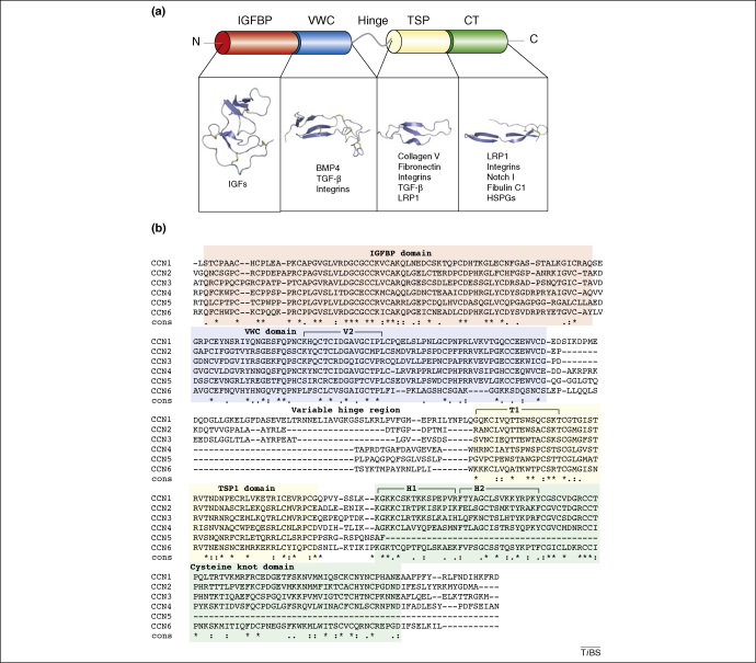
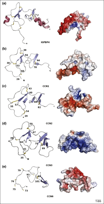
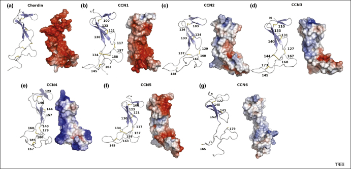
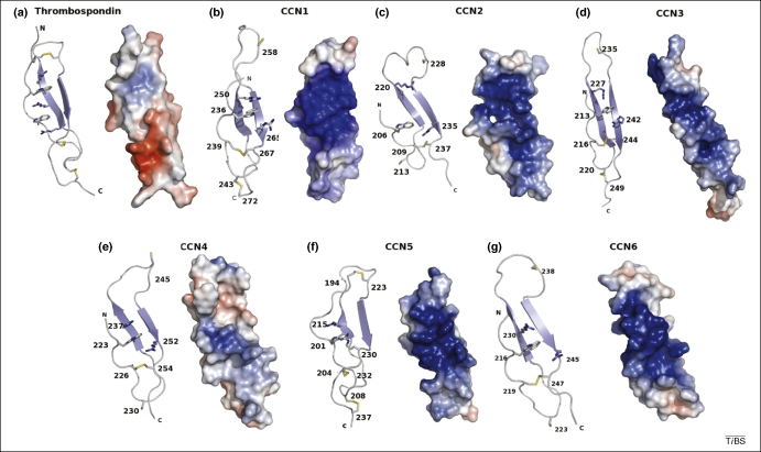
The CCN proteins are key signalling and regulatory molecules involved in many vital biological functions, including cell proliferation, angiogenesis, tumourigenesis and wound healing. How these proteins influence such a range of functions remains incompletely understood but is probably related to their discrete modular nature and a complex array of intra- and inter-molecular interactions with a variety of regulatory proteins and ligands. Although certain aspects of their biology can be attributed to the four individual modules that constitute the CCN proteins, recent results suggest that some of their biological functions require cooperation between modules. Indeed, the modular structure of CCN proteins provides important insight into their structure-function relationships.


Figures

Figure 1
Figure 1
Arrangement of CCN domains. (a) A diagram showing the signal peptide (SP), insulin-like growth factor binding domain (IGFBP) in red, von Willebrand factor C repeat (VWC) in blue, thrombospondin type-1 repeat (TSP-1) in yellow and cysteine knot (CT) in green. The protein is split into two halves separated by a variable ‘hinge’ region. Some of the known binding partners of each module are also listed: insulin-like growth factors (IGFs); bone morphogenic protein 4 (BMP4); transforming growth factor β (TGF-β); LDL receptor protein 1 (LRP-1); and heparin sulphated proteoglycans (HSPGs). (b) A sequence alignment of the CCN protein family. The sections of the sequence corresponding to each domain are shaded according to the colour scheme used in (a). The asterisks highlight the conserved residues and include the 38 cysteines that form part of the key motifs of each domain. The three regions of the sequence that have been implicated directly in integrin binding are also highlighted. These areas are highlighted in bold text. The V2 site binds integrin αvβ3 ; the T1 site binds α6β1 ; the H1 site also binds α6β1; and the H2 site binds HSPGs . The alignment was constructed by the T-Coffee server .
Figure 2
Figure 2
The IGFBP domain. The models of the IGFBP domain of (ii–iv) CCN1–3 and (v) CCN6 alongside (i) the IGFBP4 structure ([PDB code 2DSP) . The models maintain the L-shaped structure with a disulphide supported ladder-like structure forming one lobe and a β-sheet-containing section forming the second lobe. In the surface representations of the IGFBP domains red represents negative charge and blue represents positive charge. The long protruding thumb region in IGFBP4 is clearly visible but could not be modelled for any of the CCN proteins. This figure was drawn using PyMOL (http://pymol.sourceforge.net/).
Figure 3
Figure 3
The VWC domain. The ∼70 residue stretch of the VWC domain from (ii–vii) the six CCN proteins alongside (i) the structure of the VWC domain from human chordin (PDB code IU5 M) shown as ribbon and electrostatic surface models. The top half of the molecule with the two β-sheets is the first subdomain, and the lower half of the molecule held together only by disulphides is the part that resembles fibronectin and might be involved in growth factor binding. In the electrostatic surface representations of the VWC domains, positive charge is shaded blue and negative charge is shaded red. The differences in surface charge might contribute to the different biological functions observed for CCN family members. This figure was drawn using PyMOL (http://pymol.sourceforge.net/).
Figure 4
Figure 4
The CCN TSP domain. The ∼40 residue models of the TSP domain of (ii–vii) the CCN family shown alongside (i) the TSP-1 domain of thrombospondin (PDB code 1LSL) , shown as both ribbon models and electrostatic surface models. The two β-strands and the third unordered strand form the basis of the domain. Several residues that could form the ‘CWR’ layers are also shown in stick form. The first cysteine disulphide formation that forms the ‘top’ layer is not present in the model but is present in the protein sequence, and the unpaired half of it is shown at the top of the third strand. There are also two arginine residues and a tryptophan present that can form the CWR layers. The additional CWR layers in thrombospondin can also be observed. In the model structures of the TSP domains from the CCN family shown, positive charge is shaded blue and negative charge is shaded red. In each case, there is a large patch of positive charge that forms a groove that might permit interactions with heparin or other sulphated proteoglycans. This figure was drawn using PyMOL (http://pymol.sourceforge.net/).
Figure 5
Figure 5
The CT domain. The ∼50 residue partial models of (ii–iv) the CT domains of CCN1–3 and (i) the structure of BMP7 (PDB code 1LXI) . In each case the cysteine knot is visible with two disulphides forming the ring and a fifth cysteine protruding through the knot, available for either inter- or intra-molecular interactions. Electrostatic surface diagrams are also shown (red illustrates negative charge and blue illustrates positive charge) showing the subtle differences between the closely related molecules. This figure was drawn using PyMOL (http://pymol.sourceforge.net/).

References

    1. Bork P. The modular architecture of a new family of growth regulators related to connective tissue growth factor. FEBS Lett. 1993;327:125–130. - PubMed
    1. Brigstock D.R. Proposal for a unified CCN nomenclature. Mol. Pathol. 2003;56:127–128. - PMC - PubMed
    1. Lau L.F., Lam S.C. The CCN family of angiogenic regulators: the integrin connection. Exp. Cell Res. 1999;248:44–57. - PubMed
    1. Brigstock D.R. The connective tissue growth factor/cysteine-rich 61/nephroblastoma overexpressed (CCN) family. Endocr. Rev. 1999;20:189–206. - PubMed
    1. Perbal B. The CCN family of genes: a brief history. Mol. Pathol. 2001;54:103–104. - PMC - PubMed

Publication types

MeSH terms

Substances

LinkOut - more resources