. 2009 Mar;17(3):354-61.
doi: 10.1016/j.joca.2008.05.021. Epub 2008 Sep 11.

Altered functional loading causes differential effects in the subchondral bone and condylar cartilage in the temporomandibular joint from young mice

Affiliations

Altered functional loading causes differential effects in the subchondral bone and condylar cartilage in the temporomandibular joint from young mice

J Chen et al. Osteoarthritis Cartilage. 2009 Mar.

Abstract

Objective: Altered loading is an important etiological factor for temporomandibular joint (TMJ) disorders. Studies examining altered loading of the TMJ have been done in rats but the response of the TMJ to altered loading in mice is largely unknown. Therefore, due to the potential usefulness of genetically engineered mice, the goal of this study was to develop a mouse TMJ altered functional loading model.

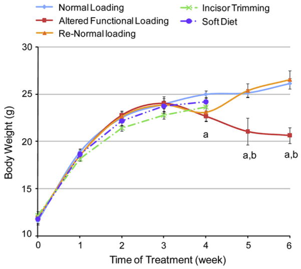
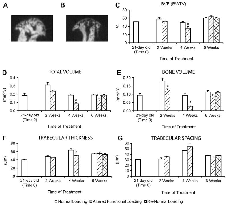
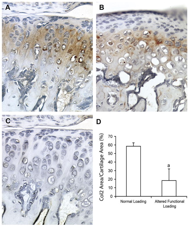
Methods: One hundred and thirty four, 21-day-old CD-1 female mice were divided into two groups: (1) normal loading (hard pellet diet) for 2-6 weeks and (2) altered functional loading (incisor trimming every other day and soft dough diet) for 2-6 weeks. The mandibular condylar cartilage was evaluated by histology, the subchondral bone was evaluated by microcomputed tomography (micro-CT) analysis and gene expression was evaluated by real time polymerase chain reaction (PCR) analysis.

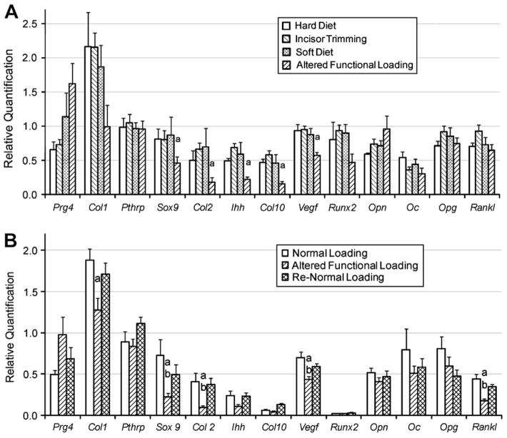
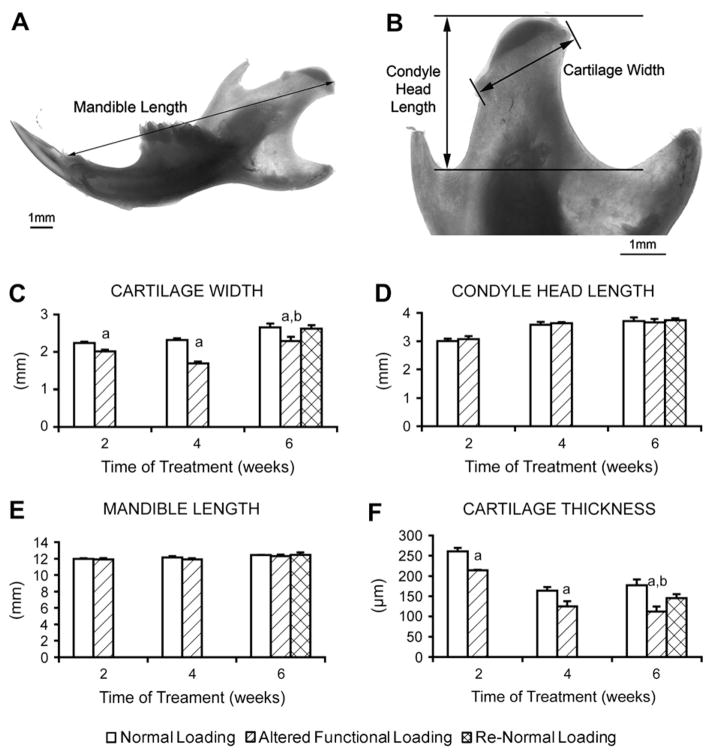
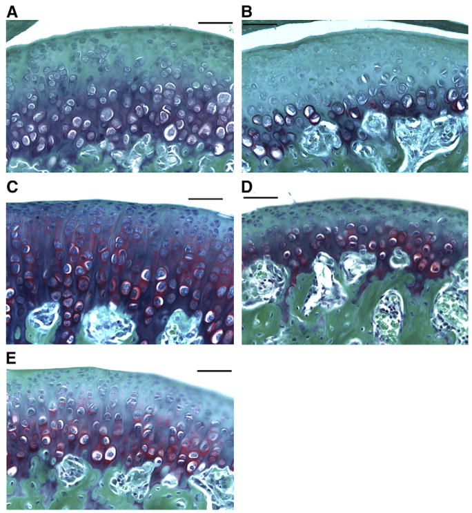
Results: Altered functional loading for 2-6 weeks caused significant reduction in the thickness of the condylar cartilage whereas, only at 4 weeks was there a significant decrease in the bone volume fraction and trabecular thickness of the subchondral bone. Gene expression analysis showed that altered functional loading for 4 weeks caused a significant reduction in the expression of SRY-box containing gene 9 (Sox9), Collagen type X (Col X), Indian hedgehog (Ihh), Collagen type II (Col II) and Vascular endothelial growth factor (Vegf) and altered loading for 6 weeks caused a significant decrease in the expression of Sox9, Col II, Vegf and Receptor activator of NF-kappaB ligand (Rankl) compared to the normal loading group.

Conclusion: Altered functional TMJ loading in mice for 2-6 weeks leads to a loss of the condylar cartilage and a transient loss in the density of the mandibular condylar subchondral bone.


Conflict of interest statement

Conflict of interest statement

All authors have no conflict of interest.

Figures

Fig. 1
Fig. 1
Body weight of female CD-1 mice exposed to different masticatory conditions. Points are the mean and S.E.M. for n = 58 for normal loading group, n = 9 incisor trimming, n = 9 soft diet, n = 59 for altered functional loading (incisor trimming in combination with soft diet administration) and n = 30 for the return to normal loading (Re-Normal). aSignificant difference between altered functional loading and normal loading (P< 0.05). bSignificant difference between altered functional loading and return to normal loading (P< 0.05).
Fig. 2
Fig. 2
21-day-old female CD-1 mice were exposed to different masticatory conditions. Real time PCR analysis for Prg4, Pthrp, Sox9, Col II, lhh, Col X, Vegf, Opn, Oc, Opg, Runx2, Col I and Rankl gene expression from the mandibular condylar head from (A) 4 weeks of normal loading, soft diet, incisor trimming or soft diet and incisor trimming (altered functional loading), and (B) 6 weeks of normal loading, altered functional loading or return to normal loading conditions (Re-Normal). Points are the mean and SEM for n = 5 for the 4-week and n = 6 for the 6-week loading groups. aSignificant difference between altered functional loading and normal loading (P < 0.05). bSignificant difference between altered functional loading and return to normal loading (P< 0.05).
Fig. 3
Fig. 3
Measurements of mandibles from 21 -day-old female CD-1 mice subjected to normal loading (2,4 or 6 weeks), altered functional loading (2, 4 or 6 weeks) and return to normal loading (Re-Normal) (6 weeks). (A and B) Gross mandibular and condylar measurements of the whole mandible and the distal condylar end of the mandible. (C—F) Statistical results of the measurements of cartilage width (C), condyle head length (D), mandible length (E) and cartilage thickness (F). Points are the mean and S.E.M. for n = 9 for the normal loading group, n = 9 for the altered functional loading group and n = 9 for the return to normal loading group. aSignificant difference between altered functional loading and normal loading (P<0.05). bSignificant difference between altered functional loading and return to normal loading (P<0.05).
Fig. 4
Fig. 4
Representatives of the Safranin O staining (red) of the condylar cartilage. The slides were counter stained with Fast Green. (A) 4-Week normal loading; (B) 4-week altered functional loading; (C) 6-week normal loading; (D) 6-week altered functional loading; and (E) return to normal loading (4-week altered loading followed by 2-week normal loading).
Fig. 5
Fig. 5
Micro-CT analysis of the subchondral bone. Representative images of mid-sagittal cross sections from the mandibular condylar head of 21-day-old CD-1 mice subjected to 4 weeks Normal Loading (A) or Altered functional loading (B). (C–G) Micro-CT analysis of bone volume fraction (C), total volume (D), bone volume (E), trabecular thickness (F) and trabecular spacing (G) from the mandibular condylar subchondral bone of 21 -day-old CD-1 mice subjected to 0 weeks (n = 4), 2 weeks (n = 11), 4 weeks (n = 10), or 6 weeks (n = 19) of normal loading, altered functional loading or return to normal loading TMJ conditions. aSignificant difference between altered loading and normal loading (P < 0.05). bSignificant difference between altered loading and return to normal loading (P< 0.05).
Fig. 6
Fig. 6
Immunohistochemistry of Col II (A – normal loading, B – altered functional loading) and negative control (C) performed on sagittal sections of the TMJ area from female CD-1 49-day-old mice. (D) Semiquantitative analysis of COL II positive area normalized by total mandibular condylar cartilage area from 49-day-old CD-1 mice exposed to 4 weeks of normal loading or altered loading. Points are the mean and SEM for n= 11 for the normal loading group and n= 11 for the altered loading group. aSignificant difference between altered loading and normal loading (P<0.05).

References

    1. LeResche L. Epidemiology of temporomandibular disorders: implications for the investigation of etiologic factors. Crit Rev Oral Biol Med. 1997;8:291–305. - PubMed
    1. Emshoff R, Brandlmaier I, Gerhard S, Strobl H, Bertram S, Rudisch A. Magnetic resonance imaging predictors of temporomandibular joint pain. J Am Dent Assoc. 2003;134:705–14. - PubMed
    1. Ohno S, Schmid T, Tanne Y, Kamiya T, Honda K, Ohno-Nakahara M, et al. Expression of superficial zone protein in mandibular condyle cartilage. Osteoarthritis Cartilage. 2006;14:807–13. - PMC - PubMed
    1. Luder HU, Leblond CP, von der Mark K. Cellular stages in cartilage formation as revealed by morphometry, radioautography and type II collagen immunostaining of the mandibular condyle from weanling rats. Am J Anat. 1988;182:197–214. - PubMed
    1. Shibukawa Y, Young B, Wu C, Yamada S, Long F, Pacifici M, et al. Temporomandibular joint formation and condyle growth require Indian hedgehog signaling. Dev Dyn. 2007;236:426–34. - PubMed

Publication types

MeSH terms