. 2008 Nov 14;383(3):667-82.
doi: 10.1016/j.jmb.2008.08.070. Epub 2008 Sep 4.

A DExH/D-box protein coordinates the two steps of splicing in a group I intron

Affiliations

A DExH/D-box protein coordinates the two steps of splicing in a group I intron

Abby L Bifano et al. J Mol Biol. .

Abstract

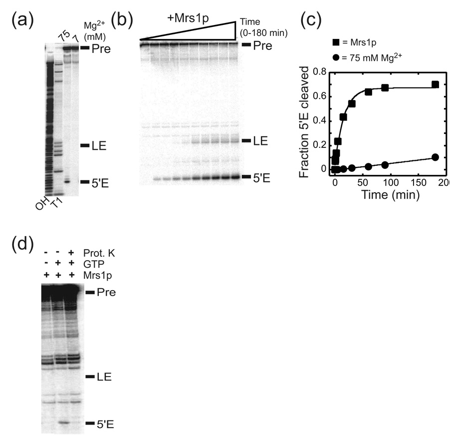
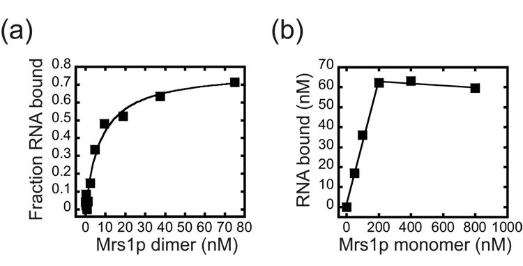
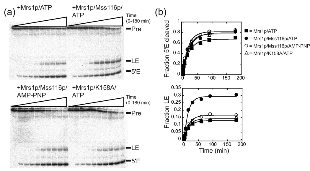
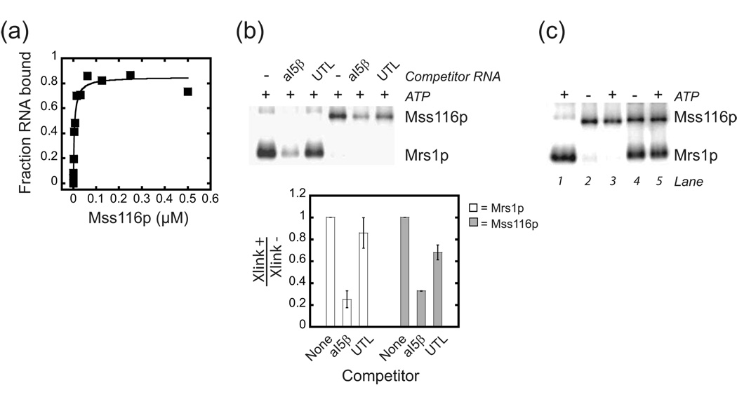
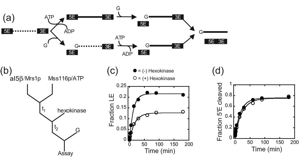
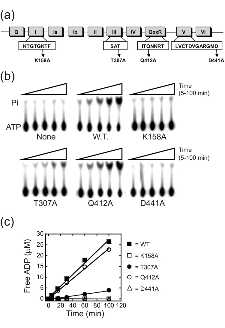
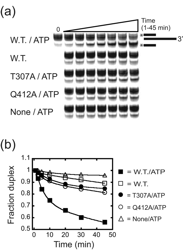
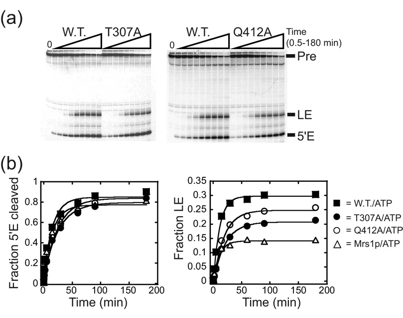
Proteins of the DExH/D family are ATPases that can unwind duplex RNA in vitro. Individual members of this family coordinate many steps in ribonucleoprotein enzyme assembly and catalysis in vivo, but it is largely unknown how the action of these co-factors is specified and precisely timed. As a first step to address this question biochemically, we describe the development of a new protein-dependent group I intron splicing system that requires such an ATPase for coordinating successive steps in splicing. While genetic analysis in yeast has shown that at least five nuclear-encoded proteins are required for splicing of the mitochondrial aI5beta group I intron, we show that efficient in vitro splicing of aI5beta occurs with only two of these co-factors and, furthermore, they fulfill distinct functions in vitro. The Mrs1p protein stabilizes RNA structure and promotes the first step in splicing. In contrast, a DExH/D protein, Mss116p, acts after the first step and, utilizing ATP hydrolysis, specifically enhances the efficiency of exon ligation. An analysis of Mss116p variants with mutations that impair its RNA-stimulated ATP hydrolysis activity or reduce its ability to unwind duplexes show that the efficiency of ATP hydrolysis is a major determinant in promoting exon ligation. These observations suggest that Mss116p acts in aI5beta splicing by catalyzing changes in the structure of the RNA/protein splicing intermediate that promote the second step. More broadly, these observations are consistent with a model in which the "functional-timing" of DExH/D-box protein action can be specified by a specific conformation of its substrate due to the "upstream" activity of other co-factors.


Figures

Figure 1
Figure 1. Splicing activity of the aI5β intron RNA
(a) Representative gel image for aI5β self-splicing reactions. 5’ 32P-end-labeled RNA was incubated in reaction buffer at 37 °C containing 75 or 7 mM MgCl2 in the presence of 1 mM GTP. Pre, aI5β pre-RNA; LE, ligated exons; 5’E, free 5’ exon; OH, hydrolysis ladder; T1, ribonuclease T1 sequencing ladder. Note that a band corresponding to ligated exons cannot be seen in the self-splicing reactions. (b) Representative gel image for Mrs1p-dependent splicing of the aI5β intron. (c) Representative plots for self-splicing and Mrs1p-dependent splicing. The total fraction of RNA that had undergone the first step (5'E cleaved) was calculated by summing the amount of radioactivity of the free 5’ exon and ligated exons and dividing this value by the total radioactivity in the lane. The values for total fraction of 5’ exon cleaved were plotted against time and the data were fit to a first order equation: Fraction RNA spliced = A(1 − ekt) where A is the amplitude of reacted RNA and k represents the pseudo-first order rate constant, kobs. See Table 1 for values and error calculations. (d) Proteinase K treatment abolishes Mrs1p stimulation of splicing. Mrs1p/aI5β pre-RNA were preincubated in the absence of GTP and the complex then digested with Proteinase K. GTP was added and the reactions proceeded for 5 min.
Figure 2
Figure 2. Binding of Mrs1p to aI5β pre-RNA
(a) Equilibrium binding. Representative plot for Mrs1p binding to aI5β RNA. The Kdapp from three independent experiments was 5.6 (± 1.7) nM. (b) Stoichiometry of the Mrs1p/aI5β pre-RNA complex. Representative plot for binding of 80 nM aI5β RNA with increasing concentrations of Mrs1p (50 – 800 nM, monomer). The reactions were conducted with RNA concentrations greater than 10-fold above the Kdapp of 5.6 nM. The plot was linear up to 200 nM Mrs1p monomers with the inverse of the slope equal to the number of protein monomers bound to the RNA. From three independent experiments, the stoichiometry of the complex was 4.3 (± 0.9) monomers per RNA.
Figure 3
Figure 3. Mss116p stimulated exon ligation
(a) Representative gel image for aI5β splicing in the presence of Mss116p and/or Mrs1p. For the wild-type Mss116p and the ATPase mutant, K158A (see Figure 6), the reactions were carried out in the presence of ATP. The non-hydrolyzable ATP analog, AMP-PNP, was also included in a set of reactions with the wild-type protein. (b) Representative plots for splicing. The fraction of RNA that had undergone the first step (5’ E cleaved) is plotted against time and the data fit to a first order equation to obtain kobs and the total fraction (amplitude) that had undergone the first step (see Figure 1). Likewise, the fraction of ligated exons was plotted against time and the data fit to a first order equation to obtain kobs and the total fraction (amplitude) that had undergone the second step. The individual amplitudes of ligated exon shown in this Figure vary less than 12% from the averaged value from multiple experiments for each condition. The average amplitudes were associated with errors of less than 20% for each condition. For values and calculated errors, see Table 1.
Figure 4
Figure 4. Interaction of Mss116p and Mrs1p with the aI5β pre-RNA
(a) Representative plot of equilibrium binding of Mss116p to aI5β pre-RNA. The Kdapp from three independent experiments was 18 (± 7.2) nM. (b) Specificity of uv cross-linking of 4-thiouridine substituted aI5β pre-RNA to Mrs1p and Mss116p. Internally 32P-radiolabeled substituted aI5β was incubated with either saturating amounts of Mrs1p (0.75 µM, dimer) or Mss116p (0.75 µM) in the absence or presence of 0.6 µM unlabeled competitor aI5β pre-RNA (aI5β) or COX3 5’ UTL (UTL). Below shows a histogram with the ratio of phosphorimager counts from cross-linked material in the presence (Xlink+) versus absence (Xlink−) of competitor RNAs. The data are from two independent experiments and the error bars represent the range of values divided by two. (c) Evidence that Mrs1p and Mss116p bind simultaneously to aI5β pre-RNA. UV cross-linking reactions were performed with saturating Mrs1p (0.75 µM, dimer, lane 1), Mss116p (0.75 µM, lanes 2,3) or both (lanes 4,5). There are no significant changes in the intensity of cross-linking when both proteins were included in the reactions suggesting that both proteins bind to a single aI5β pre-RNA. In addition, the presence of ATP does not affect the cross-linking efficiency of Mss116p or Mrs1p in the presence of Mss116p. Note that the amount of Mrs1p cross-linked is reduced ~30% in the presence of Mss116p similar to the amount reduced in the presence of the non-specific competitor COX3 UTL in (b). This may reflect that a small percentage of the Mrs1p-cross-link represents a non-specific interaction and Mss116p competes for these sites on the RNA.
Figure 5
Figure 5. Mss116p acts after the first step in splicing
(a) Schematic of possible sites of Mss116p action in the splicing cascade. Mss116p could possibly act upon precursor (top) or the intron-3’exon intermediate (bottom) RNAs to facilitate the second step. The dashed and solid lines represent structures of the intron that are incapable or capable of performing exon ligation, respectively. Removing ATP by hexokinase treatment prior to the addition of GTP (G) should abolish the increase in ligated exon, if Mss116p acts after the first step. (b) Outline of the ATP depletion protocol. Mrs1p/aI5β RNA complexes were pre-incubated with Mss116p and ATP (t1). Hexokinase was added, which catalyzes the phosphorylation of glucose via ATP hydrolysis, and, after a brief incubation (t2), splicing initiated by the addition of GTP (G). (c) Representative plot of exon accumulation. The fraction of ligated exons was plotted against time and the data fit to a single exponential to obtain kobs and the total fraction (amplitude) that had undergone the second step. The individual amplitudes of ligated exon shown in this Figure vary less than 8% from the averaged value from multiple experiments for each condition. The average amplitudes were associated with errors of less than 4% for each condition. For values and calculated errors, see Table 2. (d) Representative plots for the first step in splicing. See Table 2 for values and calculated errors.
Figure 6
Figure 6. ATPase activity of wild-type and mutant derivatives of Mss116p
(a) Schematic of Mss116p conserved motifs. Mutated residues are indicated. (b) Representative TLC plate images for each mutant in the presence of 50 µM ATP. Pi, released phosphate; None, no protein added; W.T., wild-type Mss116p. (c) Representative plots of free ADP versus time for reactions with 50 µM ATP. The curves are linear fits to the data, with the slope equal to the initial velocity (vo). For kinetic parameters, see Table 3.
Figure 7
Figure 7. Unwinding activity of wild-type and mutant derivatives of Mss116p
(a) Representative gel image for reactions in the presence of ATP. The mobility of the released, 32P-labeled DNA was confirmed by boiling the complex and running that material alongside the starting material (not shown). (b) Representative time course plots. Reactions were initiated by the addition of Mss116p, mutant protein or protein dilution buffer (None) and aliquots removed from 1 to 45 min. The data for Mss116p (W.T.), T307A and Q412A with ATP was fit to a first order equation with a double exponential: Fraction Duplex = A(ekt) + B(ekt) where A and B are the amplitude of duplex in each phase and k represents the first order rate constants for each phase. For Mss116p in the absence of ATP and in reactions without protein, the data was fit to a single exponential: Fraction Duplex = A(ekt) where A is the amplitude and k represents the first order rate constant. For values derived from multiple experiments and calculated errors, see Table 4. Multiple bands in the duplex are due to the addition of non-templated nucleotides to the RNA transcript.
Figure 8
Figure 8. Splicing activity of wild-type and mutant derivatives of Mss116p
(a) Representative gel image for time course experiments. The reactions were initiated by addition of Mrs1p and Mss116p proteins and aliquots removed from 0.5 to 180 min. (b) Representative plots of splicing product accumulation. The fraction of RNA species spliced was plotted against time and the data fit to a single exponential to obtain kobs and the total fraction (amplitude) that had undergone either step (see Figure 3). Note that the mutants stimulate the ligated exon production above that in the presence of Mrs1p alone. The individual amplitudes of ligated exon shown in this Figure for each Mss116p variant vary less than 11% from the averaged value from multiple experiments and the average amplitudes were associated with errors of less than 6%. For values and calculated errors, see Table 1.
Figure 9
Figure 9. Protein-dependent splicing of the aI5β intron
Mrs1p dimers are double green rectangles and Mss116p is a blue oval. The intron initially is structured such that it is unable to carry out either step in splicing (looped, dashed line). Binding of Mrs1p stabilizes the first step, catalytically active structure (bent, dashed line). Mss116p also binds to the starting structure of aI5β, but initially, has no effect on intron activity. After binding of guanosine (G) and first step catalysis, Mss116p, in an ATP hydrolysis dependent manner, facilitates a second conformational change (dashed to solid line) in the intron to promote exon ligation. Mss116p may also facilitate release of the ligated exons, thus preventing reversal of the second step and increase the efficiency of ligation. It may also disrupt the Mrs1p interaction with the intron (not shown, but see text).

Similar articles

Cited by

References

    1. Caprara MG, Nilsen TW. RNA: versatility in form and function. Nat Struct Biol. 2000;7:831–833. - PubMed
    1. Schroeder R, Grossberger R, Pichler A, Waldsich C. RNA folding in vivo. Curr Opin Struct Biol. 2002;12:296–300. - PubMed
    1. Schroeder R, Barta A, Semrad K. Strategies for RNA folding and assembly. Nat Rev Mol Cell Biol. 2004;5:908–919. - PubMed
    1. Bokinsky G, Nivon LG, Liu S, Chai G, Hong M, Weeks KM, Zhuang X. Two distinct binding modes of a protein cofactor with its target RNA. J Mol Biol. 2006;361:771–784. - PMC - PubMed
    1. Caprara MG, Chatterjee P, Solem A, Brady-Passerini KL, Kaspar BJ. An allosteric-feedback mechanism for protein-assisted group I intron splicing. RNA. 2007;13:211–222. - PMC - PubMed

Publication types

MeSH terms