. 2008 Nov 7;283(45):30522-30.
doi: 10.1074/jbc.M805242200. Epub 2008 Sep 11.

OSTM1 bone defect reveals an intercellular hematopoietic crosstalk

Affiliations

OSTM1 bone defect reveals an intercellular hematopoietic crosstalk

Monica Pata et al. J Biol Chem. .

Abstract

The most severe form of bone autosomal recessive osteopetrosis both in humans and in the gray-lethal (gl/gl) mouse is caused by mutations in the Ostm1 gene. Although osteopetrosis is usually associated with a defect in the hematopoietic-derived osteoclast cells, this study determined that Ostm1 is expressed in many hematopoietic cells of the myeloid and lymphoid B- and T-lineages. Hematopoiesis in gl/gl mice is characterized by a marked expansion of the osteoclast lineage but also by deregulation of the lymphoid lineages with a decrease in B-lymphoid cell populations and altered distribution in T-lymphoid double and single CD4 CD8-positive cells. In committed gl/gl osteoclasts, specific Ostm1 transgene targeting showed a requirement of additional factors and/or cells for normal osteoclast function, and importantly, defined the gl osteopetrotic defect as non-cell autonomous. By contrast, gl/gl osteoclast, B- and T-lymphoid lineage phenotypes were rescued when Ostm1 is expressed under PU.1 regulation from a bacterial artificial chromosome transgene, which established an essential role for Ostm1 in hematopoietic cells in addition to osteoclasts. Together these experiments are the first to demonstrate the existence of hematopoietic crosstalk for the production of functional and active osteoclasts.


Figures

FIGURE 1.
FIGURE 1.
Ostm1 expression in hematopoietic cells. Semi-quantitative Ostm1 expression from enriched hematopoietic cells detected high levels in B-lymphocyte (B), natural killer (NK), mast (M) cells and lower expression in T-lymphocyte (T) and activated macrophage (MAC) in comparison to positive brain tissue control (C+), negative control (C), and normalized to β-actin.
FIGURE 2.
FIGURE 2.
Production, expression, and analysis of TRAP-Ostm1 transgenic mice. A, schematic representation of the transgene used to generate TRAP-Ostm1 mice. The TRAP regulatory sequences (1.3 kb), transcription initiation site (+1), first exon (500 bp), and intron 1 were cloned upstream of Ostm1 (1.055 kb) open reading frame followed by the 3′ and poly(A) of human growth hormone gene (hGH; 2.1kb). B, transgene expression in OCLs (TR) from each TRAP-Ostm1 transgenic lines was compared with non-transgenic OCL controls (NT). Endogenous Trap, Ostm1, and β-actin expression were used as controls. C, persistence of osteopetrosis was observed in homozygous gl/gl TRAP-Ostm1 transgenic mice (gl/gl TR) similar to gl/gl non-transgenic littermate (gl/gl NT) in contrast to wild-type control (+/+) as detected by x-ray analysis (top panels) and on histological bone sections (bottom panels, magnification, ×50).
FIGURE 3.
FIGURE 3.
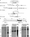
Production and analysis of BAC PU.1-Ostm1 transgene. A, schematic representation illustrates the BAC PU.1 ΔTbp1 to the BAC PU.1-Ostm1 recombination step. The PU.1 genomic sequences in BAC PU.1 ΔTbp1 were replaced by Ostm1-EGFP-poly(A) with pLD53-Ostm1-PA vector containing 5′ and 3′ PU.1 homology cassettes on each side of Ostm1-EGFP-pA. The BAC cointegrant recombination removed the pLD53.SC1 vector and produced the new BAC PU.1-Ostm1. B, the BAC PU.1-Ostm1 was used to generate transgenic lines. Southern blot of genomic DNA analysis with the following restriction digests are shown: left panel, BamHI (B) digest hybridized with a 5′ probe that detect a 3.9-kb band for the transgene; middle and right panels, EcoRI (E) or HindIII (H) hybridized with an Ostm1 probe (exon1–exon6) with one band at 7.9 kb for EcoRI digestion and two bands at 7.1 kb and 0.9 kb for HindIII digests for the transgene. Genomic DNA from the original BAC PU.1-Ostm1 was used as control.
FIGURE 4.
FIGURE 4.
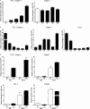
Expression analysis of the PU. 1-Ostm1 transgene. A, real-time quantitative spleen PU.1-Ostm1 transgene and endogenous Ostm1 expression from the four PU.1-Ostm1 transgenic lines (TR) and non-transgenic (NT) was established relative to S16 as internal control. B, real-time quantitative PU.1-Ostm1 transgene and Ostm1, PU.1 endogenous genes expression was carried out on different tissues of PU.1-Ostm1 transgenic (TR) mice line #761 (n = 3) relative to S16 as internal control. High expression was detected in bone marrow (BM), spleen (Sp), and thymus (Th) and lower levels in brain (Br), liver (Li), and kidney (Ki). C, real-time quantitative transgene expression was performed on bone marrow (BM) and osteoclast like cells (OCLs) generated in culture from PU.1-Ostm1 transgenic #737 (TR) and non-transgenic (NT) mice (n = 3) relative to S16 as internal control and compared with endogenous expression of Ostm1, PU.1, and TRAP.
FIGURE 5.
FIGURE 5.
Complementation of osteopetrosis in gl/gl PU. 1-Ostm1 transgenic mice. Rescue of osteopetrosis in gl/gl PU.1-Ostm1 transgenic (gl/gl TR) relative to non-transgenic (gl/gl NT) mice as shown by tooth eruption (top panels) and normal development of medullary space on bone sections stained with H&E (middle and bottom panels).

Similar articles

Cited by

References

    1. Teitelbaum, S. L., and Ross, P. (2003) Nat. Rev. Genet. 4 638–649 - PubMed
    1. Boyle, W. J., Simonet, W. S., and Lacey, D. L. (2003) Nature 423 337–342 - PubMed
    1. Zaidi, M. (2007) Nat. Med. 13 791–801 - PubMed
    1. Janssens, K., and Van Hul, W. (2002) Hum. Mol. Genet. 11 2385–2393 - PubMed
    1. Lazner, F., Gowen, M., Pavasovic, D., and Kola, I. (1999) Hum. Mol. Genet. 8 1839–1846 - PubMed

Publication types

MeSH terms