. 2008 Nov 7;283(45):31079-86.
doi: 10.1074/jbc.M806041200. Epub 2008 Sep 12.

MicroRNA-221/222 negatively regulates estrogen receptor alpha and is associated with tamoxifen resistance in breast cancer

Affiliations

MicroRNA-221/222 negatively regulates estrogen receptor alpha and is associated with tamoxifen resistance in breast cancer

Jian-Jun Zhao et al. J Biol Chem. .

Retraction in

Abstract

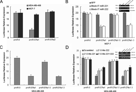
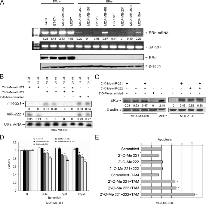
A search for regulators of estrogen receptor alpha (ERalpha) expression has yielded a set of microRNAs (miRNAs) for which expression is specifically elevated in ERalpha-negative breast cancer. Here we show distinct expression of a panel of miRNAs between ERalpha-positive and ERalpha-negative breast cancer cell lines and primary tumors. Of the elevated miRNAs in ERalpha-negative cells, miR-221 and miR-222 directly interact with the 3'-untranslated region of ERalpha. Ectopic expression of miR-221 and miR-222 in MCF-7 and T47D cells resulted in a decrease in expression of ERalpha protein but not mRNA, whereas knockdown of miR-221 and miR-222 partially restored ERalpha in ERalpha protein-negative/mRNA-positive cells. Notably, miR-221- and/or miR-222-transfected MCF-7 and T47D cells became resistant to tamoxifen compared with vector-treated cells. Furthermore, knockdown of miR-221 and/or miR-222 sensitized MDA-MB-468 cells to tamoxifen-induced cell growth arrest and apoptosis. These findings indicate that miR-221 and miR-222 play a significant role in the regulation of ERalpha expression at the protein level and could be potential targets for restoring ERalpha expression and responding to antiestrogen therapy in a subset of breast cancers.


Figures

FIGURE 1.
FIGURE 1.
Frequently increased expression of miR-221 and miR-222 in ERα-negative breast cancer. A, partial heat map of miRNA microarray analysis of ERα-positive versus ERα-negative breast cancer cell lines and primary tumors. Several miRNAs were significantly elevated in ERα-negative cells. B and C, elevated levels of miR-221 and miR-222 in ERα-negative breast cancer cell lines and primary tumors. Total RNAs from the cell lines and primary tumors were subjected to Northern blot (B) and quantitative RT-PCR (C) analyses. U6 small nuclear RNA (snRNA) and glyceraldehyde-3-phosphate dehydrogenase (GAPDH) were used as loading controls. The blots were quantified by dividing miR-221 and miR-222 signals by U6 and by dividing ERα by glyceraldehyde-3-phosphate dehydrogenase. ERα-negative tumors overexpressing both miR-221 and miR-222 are labeled by asterisks, and the tumors also expressing ERα mRNA are indicated by triangles (C). M stands for marker. D, representation of the inverse correlation of expression of ERα and miR-221/miR-222. Breast tumor specimens were immunohistochemically stained with anti-ERα antibody (first and third panels). The second and fourth panels are the same specimens that were hybridized with the LNA-miR-221 and LNA-miR-222 probes using miRNA locked nucleic acid in situ hybridization as described under “Experimental Procedures.”
FIGURE 2.
FIGURE 2.
miR-221 and miR-222 negatively regulate ERα and render cells resistant to tamoxifen. A, sequence alignment of the human miR-221 and miR-222 seed sequences with two regions of the ERα 3′-UTR. One region is conserved (ERα1; upper), and the other is not (ERα 2; lower). Mutants of pmiR-ERα1-5 (seed sequence mutation) and pmiR-ERα1-3 are shown (middle), and the mutant nucleotides are labeled in red. B, miR-221 and miR-222 inhibit ERα expression in a dose-dependent manner. MCF-7 cells were transiently transfected with indicated amounts of BLOCK-iT-miR-221 and BLOCK-iT-miR-222 expression plasmids. Following a 72-h incubation, cells were subjected to immunoblotting with anti-ERα (first panel) and β-actin (second panel) antibodies and quantitative RT-PCR analysis (third through fifth panels). Expression of transfected miR-221 and miR-222 is shown in the third and fourth panels. U6 small nuclear RNA (snRNA) is a loading control. Quantification was done by dividing ERα signals by actin. M stands for marker. C, decrease in ERα protein but not mRNA levels by stable expression of miR-221 or miR-222 in MCF-7 and T47D cells. Following transfection of BLOCK-iT-miR-221 or BLOCK-iT-miR-222, cells were selected with blasticidin. Stably transfected cells were subjected to quantitative RT-PCR (upper panels), Western blot (middle panels), and RT-PCR (lower panels) analyses. Dividing ERα signals by actin (Western) and glyceraldehyde-3-phosphate dehydrogenase (GAPDH; RT-PCR) was used to quantify the protein and mRNA levels of ERα, respectively. D, immunofluorescence staining of parental MCF-7 cells (panels A-D) and cells transiently transfected with the GFP vector (panels E-H), GFP-miR-221 (panels I-L), or GFP-miR-222 (panels M-P) with anti-ERα antibody (panels C, G, K, and O). Cells transfected with vector (e.g. expressing only GFP) exhibited the same levels of ERα as did parental cells (panels A-D). ERα signals in miR-221- and miR-222-transfected cells (arrowheads) were significantly lower than in untransfected surrounding cells (arrows). Quantitation of ERα-positive cells is shown in the bar graph. DAPI, 4′,6-diamidino-2-phenylindole. E, ectopic expression of miR-221 and miR-222 reduces tamoxifen-induced cell death. MCF-7 and T47D cells were stably transfected with miR-221, miR-222, and the vector alone as described for C. Following treatment with or without tamoxifen, cell viability was examined by 3-(4,5-dimethylthiazol-2-yl)-2,5-diphenyltetrazolium bromide assay. The experiment was repeated three times in triplicate. Asterisks indicate p < 0.05. The gels show expression of transfected miR-221 and miR-222.
FIGURE 3.
FIGURE 3.
miR-221 and miR-222 interact with the conserved site of the ERα 3-UTR. A, pmiR-ERα1 but not pmiR-ERα2 reporter activity is reduced only in miR-221/222-positive MDA-MB-468 cells. The pmiR-ERα1-Luc and pmiR-ERα2-Luc plasmids were introduced into MCF-7 and MDA-MB-468 cells together with β-galactosidase. Luciferase activity was measured and normalized after a 36-h incubation. B, ectopic expression of miR-221 or miR-222 inhibits pmiR-ERα1-Luc and pmiR-ERα1-3-Luc but not seed sequence mutant pmiR-ERα1-5-Luc. MCF-7 cells were transfected with the indicated plasmids and assayed for luciferase activity after a 36-h incubation. The inset shows expression of transfected miR-221 and miR-222. M stands for marker. snRNA, small nuclear RNA. C and D, the activities of pmiR-ERα1-Luc and pmiR-ERα1-3-Luc but not seed sequence mutant pmiR-ERα1-5-Luc are reduced in MDA-MB-468 cells (C), which are partially abrogated by knockdown of miR-221/222 (D). MDA-MB-468 cells were transfected with the indicated plasmids and 2′-O-Me. Following a 36-h incubation, luciferase reporter assay was performed as described under “Experimental Procedures.” All experiments were repeated three times in triplicate. Expression of miR-221 and miR-222 is shown in the inset.
FIGURE 4.
FIGURE 4.
Knockdown of miR-221 and/or miR-222 partially restores ERα expression and tamoxifen sensitivity in ERα protein-negative/mRNA-positive cells. A, expression of ERα protein and mRNA in breast cancer cell lines. Four ERα-positive and eight ERα-negative cell lines were subjected to RT-PCR (upper panels) and Western blot (lower panels) analyses for ERα expression. B and C, partial restoration of ERα expression by knockdown of miR-221 and miR-222. MDA-MB-468 or MCF-10A cells were transfected with 2′-O-Me-anta-miR-221 and/or 2′-O-Me-anta-miR-222. After a 72-h incubation, cells were subjected to Northern (B) and Western (C) blot analyses with the indicated probes and antibodies, respectively. Quantification was performed as described for Figs. 1 and 2. D and E, knockdown of miR-221 and miR-222 sensitizes MDA-MB-468 cells to tamoxifen-induced cell death. 2′-O-Me-anta-miR-221- and/or 2′-O-Me-anta-miR-222-transfected MDA-MB-468 cells from B were treated with the indicated doses of tamoxifen (TAM). Cell survival and apoptosis were analyzed by 3-(4,5-dimethylthiazol-2-yl)-2,5-diphenyltetrazolium bromide assay (D) and using a Cell Death Detection ELISAPLUS kit (E). Each experiment was repeated three times in triplicate. Asterisks represent p < 0.05 of tamoxifen-induced cell death between miR-221- and/or miR-222-knocked down cells and scrambled 2′-O-Me-treated cells.

References

    1. Giacinti, L., Claudio, P. P., Lopez, M., and Giordano, A. (2006) Oncologist 11 1-8 - PubMed
    1. Lee, R. C., Feinbaum, R. L., and Ambros, V. (1993) Cell 75 843-854 - PubMed
    1. Pasquinelli, A. E., Reinhart, B. J., Slack, F., Martindale, M. Q., Kuroda, M. I., Maller, B., Hayward, D. C., Ball, E. E., Degnan, B., Muller, P., Spring, J., Srinivasan, A., Fishman, M., Finnerty, J., Corbo, J., Levine, M., Leahy, P., Davidson, E., and Ruvkun, G. (2000) Nature 408 86-89 - PubMed
    1. Reinhart, B. J., Slack, F. J., Basson, M., Pasquinelli, A. E., Bettinger, J. C., Rougvie, A. E., Horvitz, H. R., and Ruvkun, G. (2000) Nature 403 901-906 - PubMed
    1. Ambros, V. (2001) Cell 107 823-826 - PubMed

Publication types

MeSH terms