. 2008 Nov 21;283(47):32870-9.
doi: 10.1074/jbc.M802863200. Epub 2008 Sep 12.

Actin binding by Hip1 (huntingtin-interacting protein 1) and Hip1R (Hip1-related protein) is regulated by clathrin light chain

Affiliations

Actin binding by Hip1 (huntingtin-interacting protein 1) and Hip1R (Hip1-related protein) is regulated by clathrin light chain

Jeremy D Wilbur et al. J Biol Chem. .

Abstract

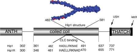
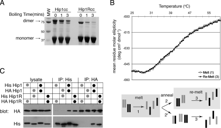
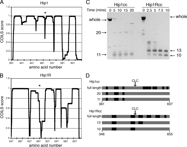
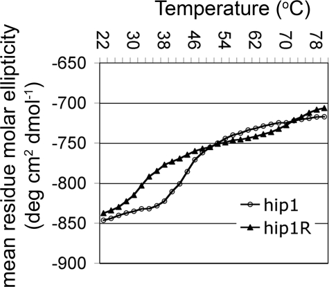
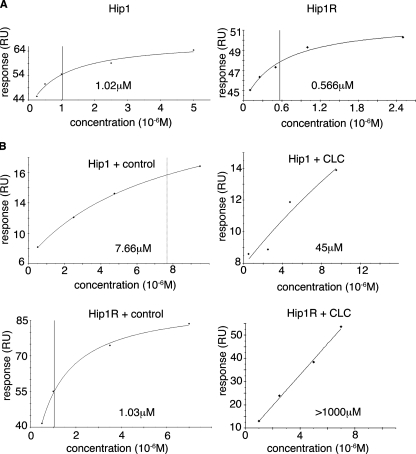
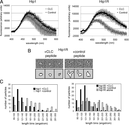
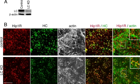
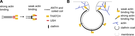
The huntingtin-interacting protein family members (Hip1 and Hip1R in mammals and Sla2p in yeast) link clathrin-mediated membrane traffic to actin cytoskeleton dynamics. Genetic data in yeast have implicated the light chain subunit of clathrin in regulating this link. To test this hypothesis, the biophysical properties of mammalian Hip1 and Hip1R and their interaction with clathrin light chain and actin were analyzed. The coiled-coil domains (clathrin light chain-binding) of Hip1 and Hip1R were found to be stable homodimers with no propensity to heterodimerize in vitro. Homodimers were also predominant in vivo, accounting for cellular segregation of Hip1 and Hip1R functions. Coiled-coil domains of Hip1 and Hip1R differed in their stability and flexibility, correlating with slightly different affinities for clathrin light chain and more markedly with effects of clathrin light chain binding on Hip protein-actin interactions. Clathrin light chain binding induced a compact conformation of both Hip1 and Hip1R and significantly reduced actin binding by their THATCH domains. Thus, clathrin is a negative regulator of Hip-actin interactions. These observations necessarily change models proposed for Hip protein function.


Figures

FIGURE 1.
FIGURE 1.
Schematic diagram of the functional domains of Hip proteins showing amino acid numbers that mark domain boundaries. The sequences of residues influencing clathrin light chain (CLC) binding regions as determined by mutagenesis are shown in alignment (3). The structure (Protein Data Bank code 2NO2) of a fragment of the Hip1 coiled-coil is shown in blue (21). Red amino acids make up the clathrin light chain binding site predicted by Ybe et al. (21). The ANTH domain binds phospholipids, and the THATCH domain binds actin, subject to intramolecular regulation by a USH (19) and a C-terminal segment (latch) (20).
FIGURE 2.
FIGURE 2.
Hip1 and Hip1R are stable homodimers. A, Hip1cc or Hip1Rcc, amino acids 361-637 and 346-655, respectively, were expressed and purified. Purified Hip proteins were diluted into reducing SDS-PAGE loading buffer and run on an SDS-polyacrylamide gel after boiling for the indicated times. The migration positions of monomers and dimers and molecular weight (MW) markers (in kDa) are indicated. B, circular dichroism melting analysis of a 1:1 mixture of Hip1cc and Hip1Rcc. Shown is the molar ellipticity versus temperature as determined by monitoring the change in helical content of proteins at 222 nm. Homodimers of Hip1 and Hip1R were mixed at a 1:1 ratio, and then a thermal denaturation profile was obtained (corresponding to melting step 1 in the diagram; open circles). The mixture of denatured proteins was allowed to anneal slowly back to 20 °C (step 2 or 2′ in the diagram). The thermal denaturation profile was collected for the newly annealed population (step 3 in the diagram; filled triangles). Both melting curves precisely overlap, showing that path 2 (not 2′) was the primary route followed. C, co-immunoprecipitation of His- or HA-tagged full-length Hip1 or Hip1R. HeLa cells were co-transfected with the two constructs indicated in the table above the blot to achieve equivalent levels of expression of each construct. HeLa cells were lysed, and tagged Hip proteins were isolated by immunoprecipitation (IP) with either anti-His antibody or anti-HA antibody as shown. Immunoprecipitates and transfected cell lysate were separated by SDS-PAGE and transferred to nitrocellulose for immunoblotting. Nitrocellulose membranes were probed with anti-His, stripped, and then probed with anti-HA. Black dots indicate homotypic transfection combinations. Gray dots indicate heterotypic transfection combinations.
FIGURE 3.
FIGURE 3.
Flexibility within the Hip1 and Hip1R coiled-coils. A and B, coiled-coil propensity of Hip1 and Hip1R coiled-coil domains, respectively, as predicted by the program COILS (34). An asterisk indicates the approximate position of residues known to be important for light chain binding, as determined by mutagenesis (3). C, partial proteolysis of Hip1 and Hip1R coiled-coil domains. 12.5 μg of Hip1 or Hip1R was digested with 0.01 μg/ml of subtilisin protease for the indicated times. Reactions were stopped by boiling in SDS-PAGE loading buffer. Proteolysis products were separated by SDS-PAGE. Fragment sizes in kDa are indicated. D, whole Hip1 or Hip1R coiled-coils or subtilisin proteolysis products indicated by arrows in C were excised from the SDS-polyacrylamide gel and further digested with trypsin. Peptides from the tryptic digests were identified by MALDI mass spectrometry. The black regions of the diagram indicate tryptic peptides identified by mass spectrometry in each digestion reaction, labeled according to the molecular weight of the band digested. Tryptic peaks missing (gray) in the subtilisin fragments indicate which regions were digested during subtilisin proteolysis. CLC marks the approximate position of clathrin light chain binding.
FIGURE 4.
FIGURE 4.
Thermal stability of Hip1 and Hip1R. Purified coiled-coil domains of Hip1 and Hip1R were subjected to increasing temperature while the change in ellipticity at 222 nm was monitored by circular dichroism. The heating rate was 0.5 °C/min. The mean residue molar ellipticity versus temperature was plotted. The transition for the thermal denaturation of Hip1 coiled-coil domain had a Tm = 43 °C (○). The thermal denaturation profile of Hip1R had a Tm = 32 °C (▴) for the first and primary transition.
FIGURE 5.
FIGURE 5.
SPR binding of Hip1 and Hip1R to clathrin light chain and actin. Representative SPR plots for steady state binding between Hip proteins and clathrin light chains or actin are shown. Fitted binding affinities are noted on each plot, with a vertical line also indicating fitted binding affinity. A, purified clathrin light b (LCb neuronal isoform) was immobilized on a CM5 chip by amine coupling. Purified coiled-coil domains of Hip1 or Hip1R at the concentrations indicated were flowed over the clathrin light chain surface until a steady state was reached, and the steady state response units (RU) were plotted. B, actin purified from rabbit muscle was assembled and immobilized on a CM5 chip by amine coupling. Purified Hip1 or Hip1R fragments containing the coiled-coil and THATCH domains (Hip1ccth or Hip1Rccth) were flowed over the F-actin surface at the concentrations indicated in the presence of saturating amounts (20 μm) of clathrin light chain peptide (CLC) or GroEL control peptide. All data were collected on a Biacore T100. BiaEvaluation software (Biacore) was used for steady state analysis of binding data. Curves with clathrin light chain peptide have no line indicating binding affinity, because fitted binding affinity was off the scale.
FIGURE 6.
FIGURE 6.
Hip1 and Hip1R change conformation upon clathrin light chain binding. A, purified Hip coiled-coils (Hip1cc and Hip1Rcc) or Hip coiled-coil plus THATCH fragments (Hip1ccth and Hip1Rccth) were incubated with ANS in the presence of clathrin light chain (CLC) or control peptide. ANS fluorescence spectra of the coiled-coils were subtracted from ANS spectra of the coiled-coil THATCH fragments. The difference between the spectra of the two fragments is plotted, indicating ANS fluorescence due to the THATCH domains alone in the presence of either peptide. B, representative electron microscopy images of Hip1Rccth in the presence of control or clathrin light chain peptide, stained with uranyl formate. Box edge, 270 Å. Top panels show micrographs, and bottom panels show outlines of protein images. See Fig. S2 for representative images of Hip1ccth after peptide binding. C, length distributions of particles of Hip1ccth and Hip1Rccth in the presence of clathrin light chain peptide, control peptide, or no peptide from images of uranyl formate-stained samples. Distribution was determined by measuring ∼100 particles for each condition. The control peptide is the clathrin light chain peptide with three mutations that abrogate Hip protein binding (4).
FIGURE 7.
FIGURE 7.
Immunofluorescence of Hip1R, actin, and clathrin following cellular depletion of clathrin light chains. HeLa cells were transfected with siRNAs targeting both clathrin light chain a (LCa) and clathrin light chain b (LCb) to knock down all clathrin light chains (LC KD) or with control (scrambled) siRNA at the same concentration and cultured for 72 h. A, HeLa cells were lysed after siRNA treatment, and protein levels were determined by immunoblotting. The indicated proteins were detected by a rabbit polyclonal antiserum against a conserved sequence shared by LCa and LCb (LC) (29) and β-actin (Sigma). Actin is shown as a loading control. B, cells treated with siRNA against clathrin light chain (LC KD) or with control siRNA were labeled for immunofluorescence with antibodies against clathrin heavy chain (HC) (green) and Hip1R (red). Actin was detected with fluorescent phalloidin and is shown in black and white images in the center panels or in green in the far right panels. Merged images of the indicated proteins are shown in the two right-hand panels of each row, with yellow indicating overlap of red and green labeling. In the far right panels, the central boxed area is magnified in the upper righthand corner. Bar, 10 μm.
FIGURE 8.
FIGURE 8.
Model of Hip protein conformations and effect on vesicle budding. A, model of the mechanism for regulation of actin binding to Hip proteins by clathrin light chain. Flexibility in the coiled-coil allows large scale bending of the coiled-coil upon clathrin light chain binding. This could reduce actin binding by the THATCH domain by direct blocking resulting from an intramolecular interaction between THATCH and a site that is exposed following clathrin light chain binding or indirectly by light chain inducing a conformational change that repositions the USH to an inhibitory conformation or a combination of both mechanisms. B, the decreased affinity of Hip1 and Hip1R for actin while bound to clathrin light chain suggests that Hip proteins do not interact with actin while incorporated into the clathrin coat. Instead, Hip proteins may interact with actin at the neck of the budding vesicle or edge of the clathrin coat, promoting development of a budding vesicle.

References

    1. Engqvist-Goldstein, A. E., and Drubin, D. G. (2003) Annu. Rev. Cell Dev. Biol. 19 287-332 - PubMed
    1. Legendre-Guillemin, V., Metzler, M., Charbonneau, M., Gan, L., Chopra, V., Philie, J., Hayden, M. R., and McPherson, P. S. (2002) J. Biol. Chem. 277 19897-19904 - PubMed
    1. Legendre-Guillemin, V., Metzler, M., Lemaire, J. F., Philie, J., Gan, L., Hayden, M. R., and McPherson, P. S. (2005) J. Biol. Chem. 280 6101-6108 - PubMed
    1. Chen, C. Y., and Brodsky, F. M. (2005) J. Biol. Chem. 280 6109-6117 - PubMed
    1. Wanker, E. E., Rovira, C., Scherzinger, E., Hasenbank, R., Walter, S., Tait, D., Colicelli, J., and Lehrach, H. (1997) Hum. Mol. Genet. 6 487-495 - PubMed

Publication types