. 2008 Sep;7(9):2609-20.
doi: 10.1158/1535-7163.MCT-07-2400.

Curcumin inhibits Akt/mammalian target of rapamycin signaling through protein phosphatase-dependent mechanism

Affiliations

Curcumin inhibits Akt/mammalian target of rapamycin signaling through protein phosphatase-dependent mechanism

Siwang Yu et al. Mol Cancer Ther. 2008 Sep.

Abstract

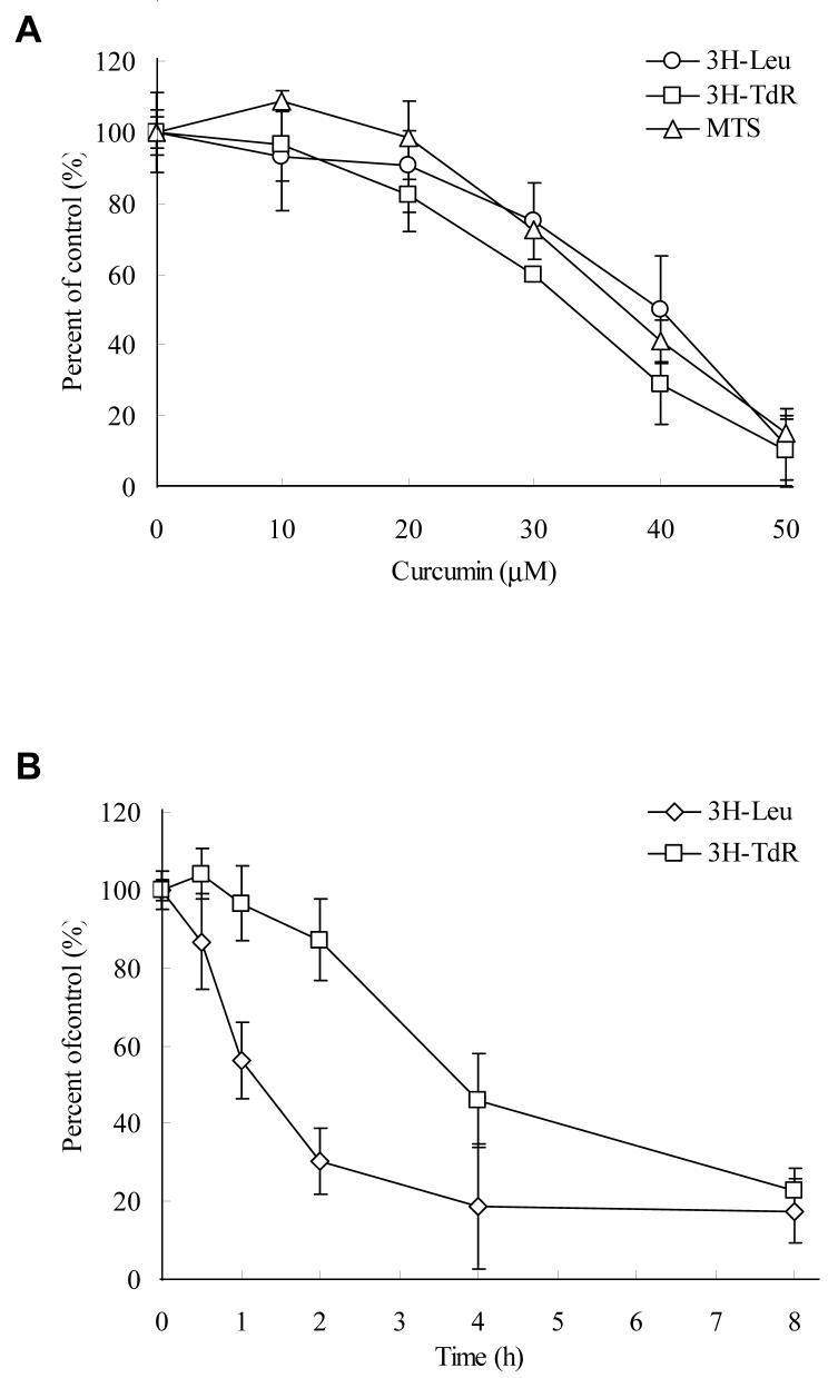
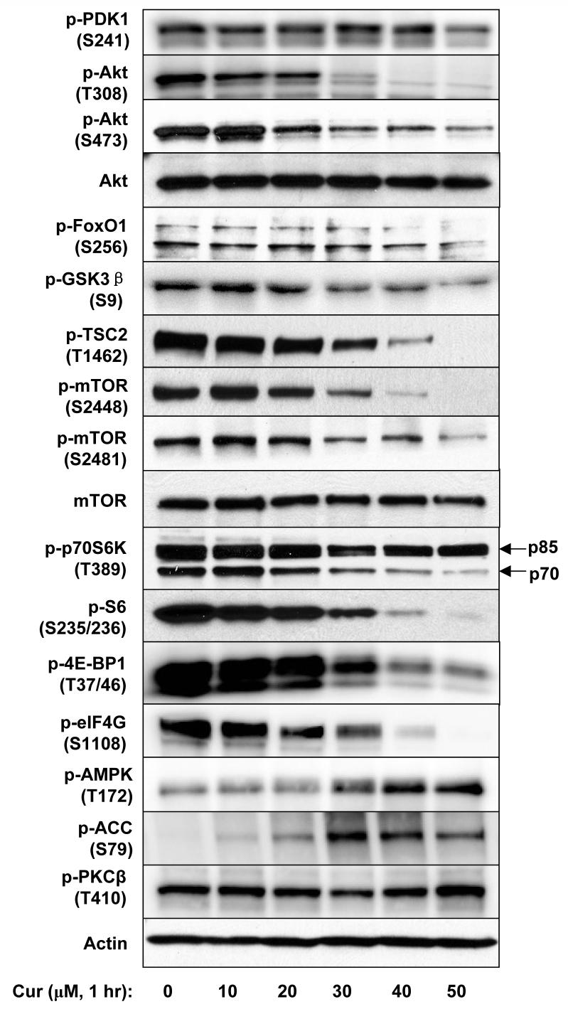
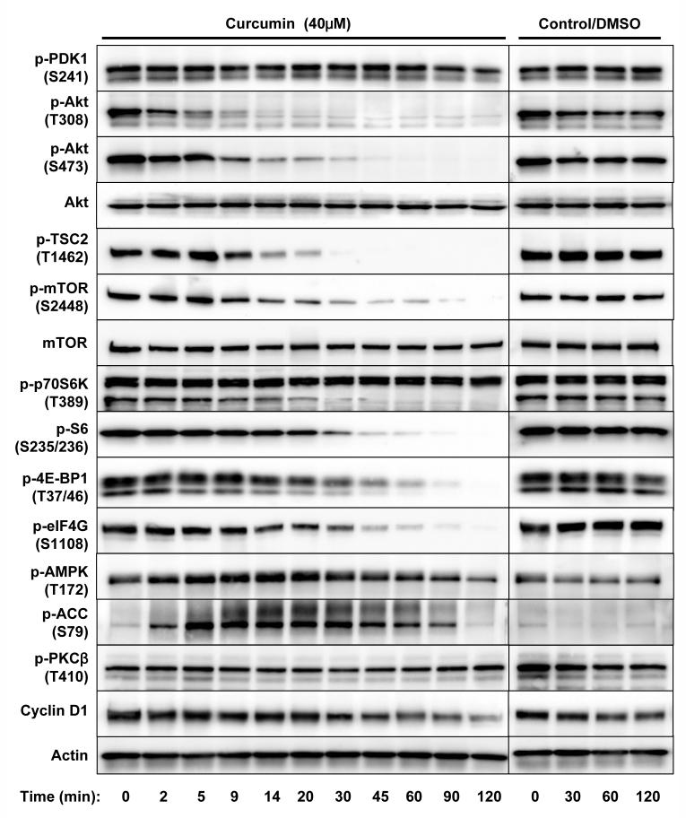
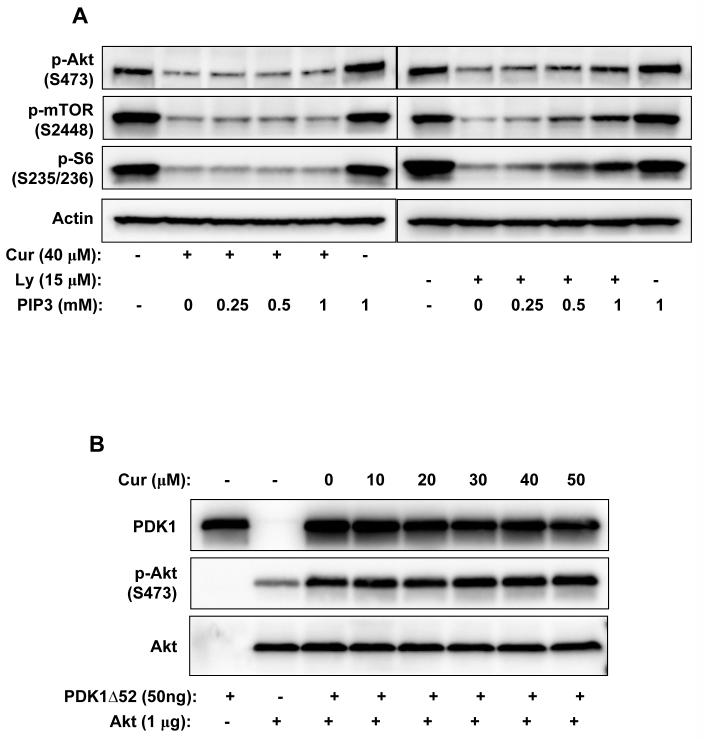
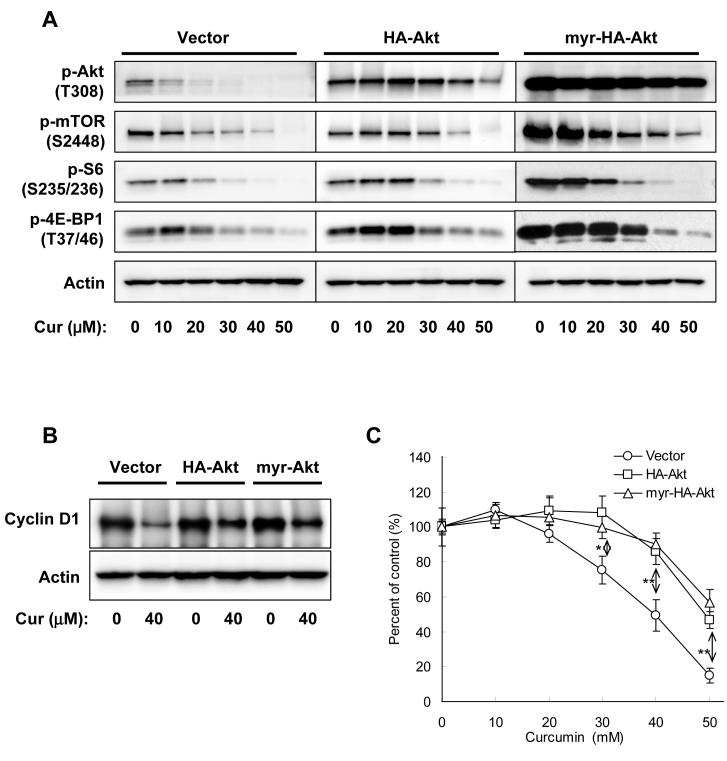
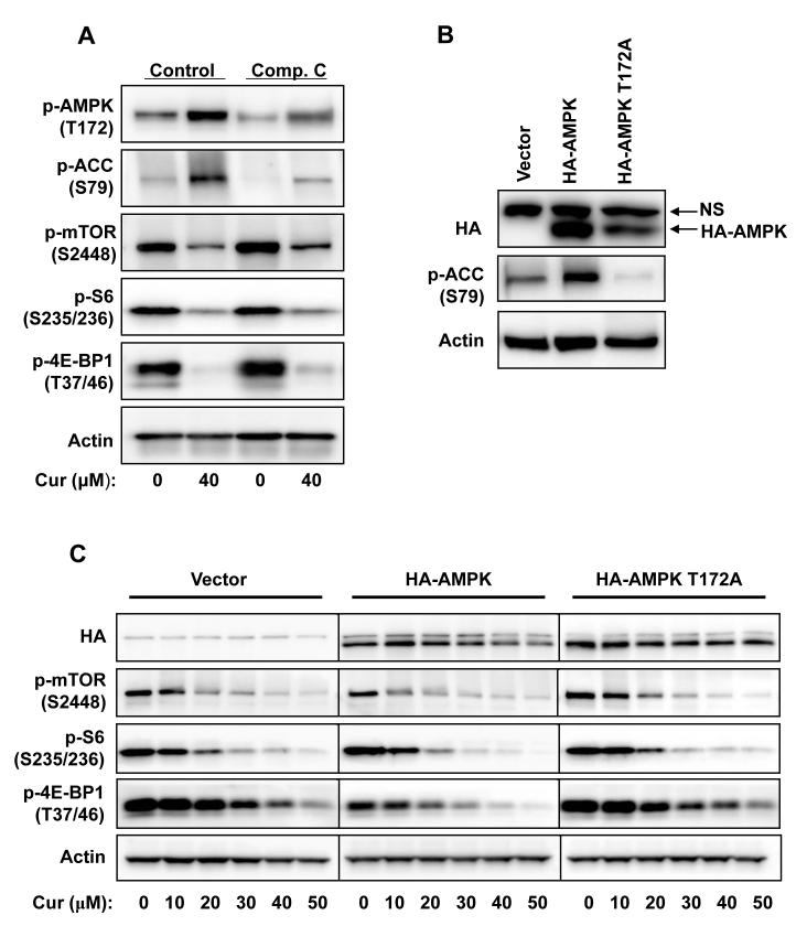
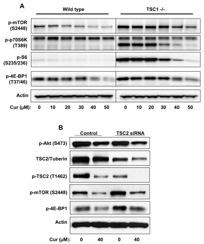
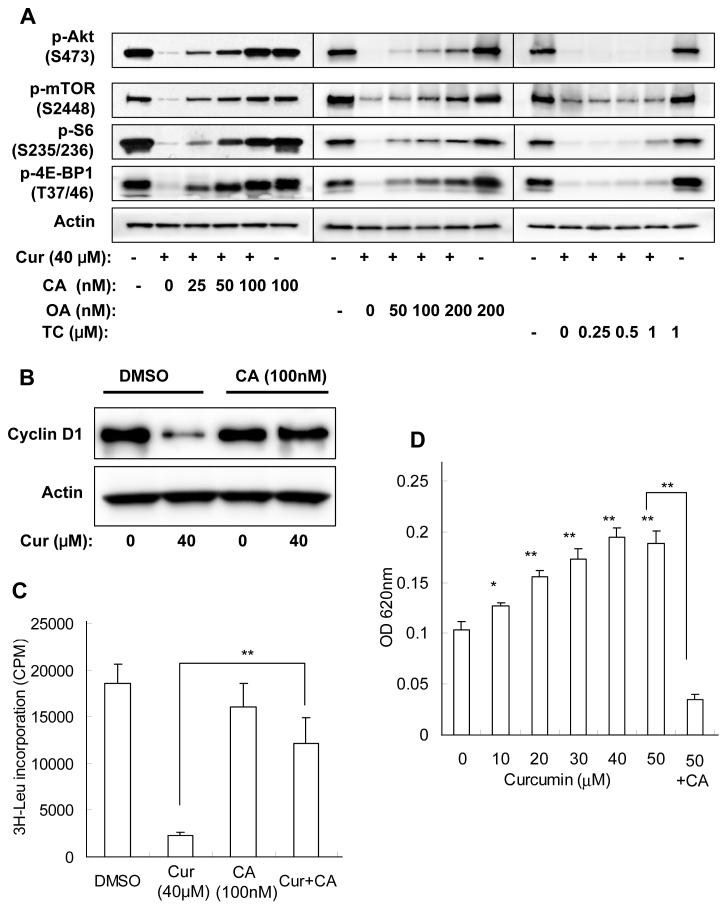
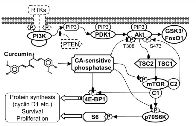
Akt/mammalian target of rapamycin (mTOR) signaling plays an important role in tumorigenesis and is dysregulated in many tumors, especially metastatic prostate cancers. Curcumin has been shown to effectively prevent or inhibit prostate cancer in vivo and inhibit Akt/mTOR signaling in vitro, but the mechanism(s) remains unclear. Here, we show that curcumin concentration- and time-dependently inhibited the phosphorylation of Akt, mTOR, and their downstream substrates in human prostate cancer PC-3 cells, and this inhibitory effect acts downstream of phosphatidylinositol 3-kinase and phosphatidylinositol-dependent kinase 1. Overexpression of constitutively activated Akt or disruption of TSC1-TSC2 complex by small interfering RNA or gene knockout only partially restored curcumin-mediated inhibition of mTOR and downstream signaling, indicating that they are not the primary effectors of curcumin-mediated inhibition of Akt/mTOR signaling. Curcumin also activated 5'-AMP-activated protein kinase and mitogen-activated protein kinases; however, inhibition of these kinases failed to rescue the inhibition by curcumin. Finally, it was shown that the inhibition of Akt/mTOR signaling by curcumin is resulted from calyculin A-sensitive protein phosphatase-dependent dephosphorylation. Our study reveals the profound effects of curcumin on the Akt/mTOR signaling network in PC-3 cells and provides new mechanisms for the anticancer effects of curcumin.


Figures

Figure 1
Figure 1
Curcumin concentration- and time-dependently inhibited cell proliferation, protein/DNA synthesis, and Akt/mTOR signaling in PC-3 cells. A, Cells were treated with various concentrations of curcumin in serum-free media for certain period then the cell viability (after 24 hrs), protein synthesis and DNA synthesis (after 8 hrs) were measured by MTS assay, 3H-Leu and 3H-TdR incorporation, respectively. B, Cells were treated with 50 μM of curcumin for indicated time, then the protein and DNA synthesis were measured by 3H-Leu and 3H-TdR incorporation, respectively. The results are presented as percentage of untreated control and each value is the mean ± SD of 4 parallel samples. C&D, Cells were treated with indicated concentrations of curcumin or DMSO (control) in serum-free media for indicated time, then harvested and immuno-blotted against indicated proteins using their phosphor-specific antibodies. Actin was blotted as a loading control. Please note that two isoforms of S6K were visualized but the p85 isoform was not affected by curcumin.
Figure 1
Figure 1
Curcumin concentration- and time-dependently inhibited cell proliferation, protein/DNA synthesis, and Akt/mTOR signaling in PC-3 cells. A, Cells were treated with various concentrations of curcumin in serum-free media for certain period then the cell viability (after 24 hrs), protein synthesis and DNA synthesis (after 8 hrs) were measured by MTS assay, 3H-Leu and 3H-TdR incorporation, respectively. B, Cells were treated with 50 μM of curcumin for indicated time, then the protein and DNA synthesis were measured by 3H-Leu and 3H-TdR incorporation, respectively. The results are presented as percentage of untreated control and each value is the mean ± SD of 4 parallel samples. C&D, Cells were treated with indicated concentrations of curcumin or DMSO (control) in serum-free media for indicated time, then harvested and immuno-blotted against indicated proteins using their phosphor-specific antibodies. Actin was blotted as a loading control. Please note that two isoforms of S6K were visualized but the p85 isoform was not affected by curcumin.
Figure 2
Figure 2
A, PIP3 rescued PI3K inhibitor LY294002-mediated but not curcumin-mediated inhibition of Akt/mTOR signaling in PC-3 cells. Cells were incubated with 40μM of curcumin or 15μM of LY294002 in the presence of increasing concentrations of PIP3 for 1 hr, then harvested and the indicated proteins were blotted. B, Curcumin did not inhibit PDK1 activity towards Akt. 1 μg of purified His-tagged Akt1 protein was incubated with 50 ng of purified active PDK1 (PDK1Δ52) in the presence of indicated concentrations of curcumin in kinase assay buffer/1 mM ATP for 20 min, then phosphor-Akt T308 and PDK1 were immuno-blotted.
Figure 3
Figure 3
Overexpression of HA-Akt or myr-HA-Akt only partially rescued curcumin-mediated inhibition in PC-3 cells. A, Cells were transfected with equal amount of indicated plasmids for 24 hrs, serum-starved for 12 hrs, then treated with various concentrations of curcumin in serum-free media for 1 hr, and harvested and blotted against indicated proteins. B, Cells were transfected with equal amount of indicated plasmids for 24 hrs, then treated with 40 μM of curcumin in serum-free media for 4 hrs, and harvested and blotted against cyclin D1 or actin. C, Cells were transfected with equal amount of indicated plasmids for 24 hrs, and then treated with various concentrations of curcumin in serum-free media for 24 hrs, and the cell viability was determined by MTS assay. The results are presented as percentage of untreated control and each value is the mean ± SD of 4 parallel samples. The difference between vector and HA-Akt or myr-HA-Akt is verified by t test. *, p < 0.05; **, p < 0.01.
Figure 4
Figure 4
Activation of AMPK is not the major reason for curcumin-mediated inhibition of mTOR signaling in PC-3 cells. A, Cells were pretreated with 10 μM of compound C for 15 min then treated with 40μM of curcumin in the presence of compound C for 1 hr, then harvested and blotted against indicated proteins. B, Cells were transfected with indicated plasmids for 24 hrs, then harvested and blotted against HA, phosphor-ACC and actin. NS: Non-specific band. C, Cells were transfected with indicated plasmids for 24 hrs, serum-starved for 12 hrs and then treated with various concentrations of curcumin in serum-free media for 1 hr, and harvested and blotted against indicated proteins.
Figure 5
Figure 5
Disruption of TSC1-TSC2 complex failed to rescue curcumin-mediated inhibition of mTOR signaling. A, Wild type or TSC1 (-/-) MEFs were serum-starved for 12 hrs then incubated with various concentrations of curcumin in serum-free media for 1 hr, and harvested and blotted against indicated proteins. B, PC-3 cells were transfected with siRNA against TSC2/tuberin or scrambled control RNA for 48hrs, serum-starved for 12hrs, then treated with 40 μM of curcumin in serum-free media for 1 hr, and harvested and blotted against proteins indicated.
Figure 6
Figure 6
Curcumin-mediated inhibition of Akt/mTOR signaling in PC-3 cells is dependent on PP2A and/or unspecified calyculin A-sensitive protein phosphatase activity. A, Cells were pretreated with indicated concentrations of protein phosphatase inhibitors for 15 min, then incubated with 40 μM of curcumin in the presence of protein phosphatase inhibitors in serum-free media for 1 hr, and harvested and blotted against indicated proteins. B&C, Cells were pretreated with 100 nM of calyculin A for15 min, then incubated with 40 μM of curcumin in the presence of calyculin A for 4 hrs, and then (B) cells were harvested and blotted against cyclin D1 and actin or (C) 3H-Leu incorporation was determined as described in Material and Methods. D, Cells were treated with indicated concentrations of curcumin for 10 min, then harvested in phosphatase lysis buffer and the protein phosphatase activities in lysates were determined as described in Material and Methods. The results are expressed as mean ± SD of 4 parallel samples. *, p < 0.05; **, p < 0.01.
Figure 7
Figure 7
Summary of the mechanisms by which curcumin inhibits Akt/mTOR signaling and cell survival/proliferation in PC-3 prostate cancer cells. Curcumin activated PP2A and/or unspecified calyculin A-sensitive protein phosphatase activities towards Akt, mTOR, led to the dephosphorylation of Akt/mTOR and their downstream substrates GSK3, FoxO1, p70S6K and 4E-BP1, and finally inhibited the expression of proteins that are essential for cell survival and proliferation. Curcumin also activated MAPKs and AMPK; however these kinases did not play important roles in the curcumin-mediated inhibition of Akt/mTOR signaling and cell proliferation.

Similar articles

Cited by

References

    1. Hennessy BT, Smith DL, Ram PT, Lu Y, Mills GB. Exploiting the PI3K/AKT pathway for cancer drug discovery. Nat Rev Drug Discov. 2005;4:988–1004. - PubMed
    1. Sarbassov DD, Guertin DA, Ali SM, Sabatini DM. Phosphorylation and regulation of Akt/PKB by the rictor-mTOR complex. Science. 2005;307:1098–101. - PubMed
    1. Belham C, Wu S, Avruch J. Intracellular signalling: PDK1--a kinase at the hub of things. Curr Biol. 1999;9:R93–6. - PubMed
    1. Toker A, Yoeli-Lerner M. Akt signaling and cancer: surviving but not moving on. Cancer Res. 2006;66:3963–6. - PubMed
    1. Cully M, You H, Levine AJ, Mak TW. Beyond PTEN mutations: the PI3K pathway as an integrator of multiple inputs during tumorigenesis. Nat Rev Cancer. 2006;6:184–92. - PubMed

Publication types

MeSH terms

Substances