. 2008 Nov;131(Pt 11):3081-91.
doi: 10.1093/brain/awn217. Epub 2008 Sep 12.

Identification of soluble TREM-2 in the cerebrospinal fluid and its association with multiple sclerosis and CNS inflammation

Affiliations

Identification of soluble TREM-2 in the cerebrospinal fluid and its association with multiple sclerosis and CNS inflammation

Laura Piccio et al. Brain. 2008 Nov.

Abstract

Triggering receptor expressed on myeloid cells 2 (TREM-2) is a membrane-bound receptor expressed by microglia and macrophages. Engagement of TREM-2 on these cells has been reported to reduce inflammatory responses and, in microglial cells, to promote phagocytosis. TREM-2 function is critical within the CNS, as its genetic deficiency in humans causes neurodegeneration with myelin and axonal loss. Blockade of TREM-2 worsened the mouse model for multiple sclerosis. In the present study, a soluble form of TREM-2 protein has been identified by immunoprecipitation and by ELISA. Soluble TREM-2 protein (sTREM-2) was detected in human CSF, and was compared among subjects with relapsing-remitting multiple sclerosis (RR-MS; n = 52), primary progressive multiple sclerosis (PP-MS; n = 21), other inflammatory neurologic diseases (OIND; n = 19), and non-inflammatory neurologic diseases (NIND; n = 41). Compared to NIND subjects, CSF sTREM-2 levels were significantly higher in RR-MS (P = 0.004 by ANOVA) and PP-MS (P < 0.001) subjects, as well as in OIND (P < 0.001) subjects. In contrast, levels of sTREM-2 in blood did not differ among the groups. Furthermore, TREM-2 was detected on a subset of CSF monocytes by flow cytometry, and was also highly expressed on myelin-laden macrophages in eight active demyelinating lesions from four autopsied multiple sclerosis subjects. The elevated levels of sTREM-2 in CSF of multiple sclerosis patients may inhibit the anti-inflammatory function of the membrane-bound receptor suggesting sTREM-2 to be a possible target for future therapies.


Figures
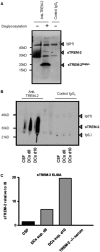
Fig. 1
Fig. 1
Soluble TREM-2 glycoprotein detected in human cerebrospinal fluid (CSF) and cultured DCs supernatant fluid. (A) Soluble TREM-2 protein (sTREM-2) was immunoprecipitated in DC supernatant with anti-TREM-2 mAbs (first and second lanes from left) or mouse IgG1 isotype control antibody (right lane). Immunoprecipitates with the anti-TREM-2 mAb were left untreated or deglycosylated to remove N- and O-linked carbohydrates. After deglycosylation (second lane) a sTREM-2 protein is detectable as a single 20 kDa band. (B) sTREM-2 glycoprotein was identified in human CSF (from a NIND subject with chronic headache) following immunoprecipitation with anti-TREM-2 mAb (first lane from left). In the supernatants of human monocyte-derived DCs after 6 or 10 days in culture, the amount of sTREM-2 protein increased with time (second and third lanes). In A and B the bands at 25 and 50 kDa represent the light (Ig L) and heavy (Ig H) chains of Ig, respectively. Molecular weight markers and specific protein bands are indicated. (C) Levels of sTREM-2 in the same specimens tested in B were quantitated by ELISA with results that were consistent with the qualitative levels observed in the immunoprecipitations. sTREM-2 was undetectable in the serum of a patient with a TREM-2 gene mutation (TREM-2–/–) leading to a truncated protein. sTREM-2 values are reported as the ratio between the samples and a positive IS.
Fig. 2
Fig. 2
Elevated levels of soluble TREM-2 in the CSF of multiple sclerosis and OIND subjects. (A) sTREM-2 in CSF samples obtained from NIND (n = 41), RR-MS (n = 52), PP-MS (n = 21) and OIND (n = 19) subjects was quantitated by ELISA. sTREM-2 levels in the CSF were significantly higher in RR-MS and PP-MS subjects compared to subjects with NIND (P = 0.004 and P < 0.001, respectively). OIND subjects also displayed CSF sTREM-2 levels significantly higher than NIND subjects (P < 0.001). (B) sTREM-2 levels assayed in the sera of RR-MS (n = 44), PP-MS (n = 19), NIND (n = 30) and OIND (n = 10) subjects showed no significant differences among groups. TREM-2 values are given as a ratio to a positive IS (a single batch of human serum positive for sTREM-2) used in all assays. The horizontal lines indicate the medians. P-values were calculated using ANOVA.
Fig. 3
Fig. 3
TREM-2 receptor is expressed on cerebrospinal fluid monocytes but not on blood monocytes. (A) TREM-2 receptor expression was evaluated on CSF cells isolated from multiple sclerosis and non-multiple sclerosis subjects. Flow cytometry staining for TREM-2 and the monocyte marker CD14 showed that TREM-2 is expressed on CSF monocytes. A plot for one representative subject from each group is shown in this figure. Sixty one percent of CSF CD14+ monocytes were also positive for TREM-2 in subject 1 (NIND, degenerative disk disease), whereas 0% of CSF CD14+ cells were TREM-2+ in subjects 2 (RR-MS) and 3 (OIND-viral meningitis). TREM-2 was never detected on blood CD14+ monocytes. (B) CSF CD14+ monocytes were positive for CD16 and CCR5 expression. Subjects 4 and 5 shown in B belonged to the NIND group. For A and B, forward vs. side scatter plots were used to gate on lymphocytes and monocytes. Dead cells were excluded by gating on propidium iodide-negative cells. In A, the percentages indicated in the upper right quadrants were calculated for each specimen as the ratio between CD14+ TREM-2+ monocytes and the total number of CD14+ monocytes.
Fig. 4
Fig. 4
Quantitation of TREM-2 receptor expression on CSF monocytes in the NIND, multiple sclerosis and OIND groups. The percentages of CD14+ CSF monocytes that were also TREM-2+ were calculated for 29 NIND subjects, 13 multiple sclerosis subjects and six OIND subjects. The percentage of CD14+ CSF monocytes dually expressing TREM-2 was significantly higher in subjects with NIND compared with multiple sclerosis subjects (P < 0.01, Kruskal–Wallis test). TREM-2 was detectable on CSF monocytes in 31% of multiple sclerosis subjects versus 83% of subjects with NIND (P = 0.003, Fisher's exact test). The horizontal lines indicate the medians.
Fig. 5
Fig. 5
TREM-2 is highly expressed on foamy macrophages in actively demyelinating multiple sclerosis lesions. (AF) Active multiple sclerosis lesion in the pons of a 46-year-old PP-MS subject characterized by the presence of perivascular inflammatory infiltrates, demyelination and numerous myelin-laden ‘foamy’ macrophages. (AC) are stained with Luxol fast blue/PAS+ and (DF) are stained with Oil Red O (ORO). Arrows in A and D denote an active multiple sclerosis lesion shown at higher magnification in B, C, E and F. (C) Mononuclear inflammatory cells forming a perivascular cuff, also seen in the upper left of B (boxed region). (E) Numerous ORO+ myelin-laden ‘foamy’ macrophages, shown at higher magnification in F. (G) Region shown in E has been immunostained for CD11b (green), TREM-2 (red) or with isotype control antibodies (shown in inset on row G). TREM-2 was highly expressed on CD11b positive lipid-laden macrophages, as demonstrated in the merged fluorescent images. Original magnification: A and D 1×, B 3.2×, E 4×, C and F 20×, G 40×. Scale bars: 2 mm in A and D, 200 μm in B and E, 50 μm in C and F, 20 μm in G.

References

    1. Begum NA, Ishii K, Kurita-Taniguchi M, Tanabe M, Kobayashi M, Moriwaki Y, et al. Mycobacterium bovis BCG cell wall-specific differentially expressed genes identified by differential display and cDNA subtraction in human macrophages. Infect Immun. 2004;72:937–48. - PMC - PubMed
    1. Bouchon A, Facchetti F, Weigand MA, Colonna M. TREM-1 amplifies inflammation and is a crucial mediator of septic shock. Nature. 2001a;410:1103–7. - PubMed
    1. Bouchon A, Hernandez-Munain C, Cella M, Colonna M. A DAP12-mediated pathway regulates expression of CC chemokine receptor 7 and maturation of human dendritic cells. J Exp Med. 2001b;194:1111–22. - PMC - PubMed
    1. Boven LA, Van Meurs M, Van Zwam M, Wierenga-Wolf A, Hintzen RQ, Boot RG, et al. Myelin-laden macrophages are anti-inflammatory, consistent with foam cells in multiple sclerosis. Brain. 2006;129:517–26. - PubMed
    1. Cella M, Buonsanti C, Strader C, Kondo T, Salmaggi A, Colonna M. Impaired differentiation of osteoclasts in TREM-2-deficient individuals. J Exp Med. 2003;198:645–51. - PMC - PubMed

Publication types

MeSH terms