. 2008 Dec;95(11):5462-75.
doi: 10.1529/biophysj.108.139428. Epub 2008 Sep 12.

Dysfunctional connections between the nucleus and the actin and microtubule networks in laminopathic models

Affiliations

Dysfunctional connections between the nucleus and the actin and microtubule networks in laminopathic models

Christopher M Hale et al. Biophys J. 2008 Dec.

Abstract

Laminopathies encompass a wide array of human diseases associated to scattered mutations along LMNA, a single gene encoding A-type lamins. How such genetic alterations translate to cellular defects and generate such diverse disease phenotypes remains enigmatic. Recent work has identified nuclear envelope proteins--emerin and the linker of the nucleoskeleton and cytoskeleton (LINC) complex--which connect the nuclear lamina to the cytoskeleton. Here we quantitatively examine the composition of the nuclear envelope, as well as the architecture and functions of the cytoskeleton in cells derived from two laminopathic mouse models, including Hutchinson-Gilford progeria syndrome (Lmna(L530P/L530P)) and Emery-Dreifuss muscular dystrophy (Lmna(-/-)). Cells derived from the overtly aphenotypical model of X-linked Emery-Dreifuss muscular dystrophy (Emd(-/y)) were also included. We find that the centrosome is detached from the nucleus, preventing centrosome polarization in cells under flow--defects that are mediated by the loss of emerin from the nuclear envelope. Moreover, while basal actin and focal adhesion structure are mildly affected, RhoA activation, cell-substratum adhesion, and cytoplasmic elasticity are greatly lowered, exclusively in laminopathic models in which the LINC complex is disrupted. These results indicate a new function for emerin in cell polarization and suggest that laminopathies are not directly associated with cells' inability to polarize, but rather with cytoplasmic softening and weakened adhesion mediated by the disruption of the LINC complex across the nuclear envelope.


Figures

FIGURE 1
FIGURE 1
Immunolocalization of lamin A and LINC complex components in wild-type and laminopathic fibroblasts. Localization of nuclear lamina protein lamin A, nuclear envelope protein emerin, and LINC complex components Sun1, Sun2, Nesprin2-giant, and Nesprin 3 in wild-type and LmnaL530P/L530P (MAFs), Lmna+/+, Lmna−/−, Emd+/y, and Emd−/y (MEF) cells. Note that Golgi is stained in Sun2 photos due to antibody cross-reaction; Sun2 localizes only to the inner nuclear membrane. Scale bar, 20 μm; pairs of images are at same scale, unless otherwise indicated.
FIGURE 2
FIGURE 2
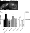
Distance between nucleus and MTOC in wild-type and laminopathic fibroblasts. (A) Typical distance between nucleus and MTOC in wild-type and LmnaL530P/ L530P MAFs. MTOC localization (open arrows) was determined by immunofluorescence microscopy using antibodies against γ-tubulin. Nuclear DNA was stained with DAPI. Scale bar, 10 μm. (B) Average distance between nucleus and MTOC in wild-type and LmnaL530P/L530P, Lmna+/+, Lmna−/−, Emd+/y, and Emd−/y cells. For each type of cell, the nucleus-MTOC distance in at least 20 cells was measured in three independent experiments (total, >60 cells per condition). **, p < 0.01; and *, p < 0.05. Asterisks shown are relative to corresponding wild-type cells (previous column). In all experiments, cells were plated on collagen.
FIGURE 3
FIGURE 3
Shear-induced polarization is abrogated in laminopathic fibroblasts. (A) Typical position of the MTOC with respect to the nucleus in a wild-type (left panel) and an LmnaL530P/ L530P (right panel) MAF, both under flow conditions. Cells were subjected to shear flow for 40 min at a wall shear stress of 20 dyn/cm2, then fixed and stained with DAPI (nuclear DNA) and an antibody against α-tubulin (α-tubulin/Alexa568), and scored for the fraction of cells with MTOCs (circled) facing away from the flow direction (i.e., located to the right of a midline through the nucleus). The flow direction is from left to right. In the absence of shear; the probability to find the MTOC at the left of the nucleus is ∼50%. Scale bar, 10 μm. (B) Average position of the MTOC with respect to the nucleus in both unsheared and sheared wild-type and LmnaL530P/L530P (MAFs), Lmna+/+, Lmna−/−, Emd+/y, and Emd−/y (MEF) cells. For each type of cell and test condition (no shear and shear), the relative position of the MTOC with respect to the nucleus in at least 20 cells was measured in at least three independent experiments (total, >60 cells per condition). *, p < 0.05. Asterisks shown are relative to corresponding wild-type cells (previous column). In all experiments, cells were plated on collagen.
FIGURE 4
FIGURE 4
Basal actin cytoskeleton architecture. (A) Typical actin filament and focal adhesion organization in wild-type and LmnaL530P/ L530P MAFs and wild-type and Lmna−/− MEFs. Cells were plated on collagen, then fixed and stained for actin (green) and focal adhesion protein vinculin (red) using phalloidin 488 and anti-vinculin/Alexa-Fluor 568, respectively. Scale bar, 20 μm. (B) Average area per focal adhesion, determined by tracing of vinculin-containing focal adhesions followed by morphometric analysis. (C) Average focal adhesion shape factor. (D) Average length of long axis of focal adhesions. (E) Average number of focal adhesions per cell. Focal adhesions were analyzed in at least 10 cells per condition, repeated in three independent experiments (total, >30 cells per condition; ∼1000 focal adhesions per condition).
FIGURE 5
FIGURE 5
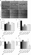
Cell migration, cell adhesion, and small GTPase activity in wild-type and laminopathic fibroblasts. (A) Typical time-dependent closure of an in vitro wound applied to wild-type (top panels) and LmnaL530P/ L530P (bottom panels) MAFs. Wound edges (shown in black as guides to the eye) were tracked by time-lapse phase-contrast microscopy over 6 h in an incubated, 5% CO2 chamber, mounted on a microscope. Scale bar, 200 μm. (B) Rate of wound healing in wild-type and LmnaL530P/L530P (MAFs), Lmna+/+, Lmna−/−, Emd+/y, and Emd−/y cells. For each condition, the rate of healing was measured in at least six different wounds whose initial width was within 50 μm of the total average wound size. (C) Cell-matrix adhesion as measured by a simple cell adhesion chamber assay (see text for details). The cell adhesion assay was performed using microtiter plates and was repeated in three independent experiments with adhesion measured in at least 36 wells per condition. (D) Average, normalized levels of activation in small GTPase RhoA. (E) Average, normalized levels of activation in small GTPase Rac. For each condition, the activation assay was performed in three independent experiments. ***, p < 0.001; **, p < 0.01; and *, p < 0.05. Asterisks shown are relative to corresponding wild-type cells (previous column). In all experiments, cells were plated on collagen.
FIGURE 6
FIGURE 6
Intracellular microrheology of wild-type and laminopathic fibroblasts. (A) Fluorescent 100-nm diameter polystyrene nanoparticles were ballistically injected in wild-type (left panel) and LmnaL530P/ L530P (right panel) MAFs. Fluorescent micrographs of nanoparticles (outlined in white circles) were superimposed on phase-contrast micrographs of cells. Representative trajectories of nanoparticles are also shown at the top right (inset) of each micrograph. Micrograph scale bar, 20 μm; inset scale bar, 0.1 μm. (B) Nanoparticles were subsequently tracked with high spatial (<10 nm) and temporal (>1/30 s) resolutions using multiparticle tracking software. Ensemble-averaged mean-squared displacements of nanoparticles embedded in the cytoplasm of wild-type (bottom black curve) and LmnaL530P/ L530P (top gray curve) MAFs are shown. (C) Mean elasticity of the cytoplasm in wild-type and LmnaL530P/L530P cells (MAFs), Lmna+/+, Lmna−/−, Emd+/y, and Emd−/y cells (MEFs). For each condition, the microrheology of at least 10 different cells was measured in three independent experiments (total, >30 cells per condition; >300 nanoparticles per condition). (D) Simplified cellular model depicting the intracellular mechanics of healthy and laminopathic cells. Healthy cells in which the nucleo-cytoskeletal connections are intact will resist force of large magnitude, while laminopathic cells cannot resist such forces as well even when actin filament architecture in these cells remains largely intact, due to defunct nucleo-cytoskeletal connections. ***, p < 0.001. Asterisks shown are relative to corresponding wild-type cells (previous column). In all experiments, cells were plated on collagen.
FIGURE 7
FIGURE 7
Localization of nuclear membrane proteins with respect to the inner and outer nuclear membranes and effects of defunct nucleo-cytoskeletal connections on cell functions. Several proteins interact across the nuclear membrane to form connections between the lamina of the inner nuclear envelope and the cellular cytoskeleton. Interactions in normal cells are shown in the figure, while the functional defects of laminopathic mutations common to both laminopathic models (LmnaL530P/L530P MAF and Lmna−/− MEF) are shown in text above the diagram. Emerin has been shown to localize at the inner nuclear membrane (55) and bind lamin A (15). A recent study also shows that emerin can localize to the outer nuclear membrane and suggests emerin's interaction with microtubules to tether the MTOC to the nucleus (37). Sun proteins localize to the inner nuclear membrane (29,30) and bind nuclear lamins in the nucleoplasmic domain (16,17), while specific Nesprin isoforms, particularly Nesprin2 giant and Nesprin3, localize to the outer nuclear membrane (31,56). Though not represented here, Sun and Nesprin proteins most likely exist as dimers (18,57,58). Sun and Nesprin proteins have been shown to interact promiscuously in the perinuclear space (45). Nesprin2 giant contains an actin-binding domain (ABD) (59), while the α-isoform of Nesprin3 binds plectin (31), a multidomain protein capable of interacting with actin, intermediate filaments, and microtubules (60). Collectively, the Sun and Nesprin proteins form the linker of nucleus and cytoskeleton, or LINC, complex, connecting the nuclear lamina to the cytoskeleton. Thus, emerin and the LINC complexes are believed to bridge connections between lamins of the inner nuclear envelope and the microtubule, actin, and intermediate filament cytoskeletal networks. (ONM, outer nuclear membrane; PNS, perinuclear space; INM, inner nuclear membrane; ABD, actin-binding domain; PBD, plectin-binding domain; and MTOC, microtubule organizing center.)

Similar articles

Cited by

References

    1. Capell, B. C., and F. S. Collins. 2006. Human laminopathies: nuclei gone genetically awry. Nat. Rev. Genet. 7:940–952. - PubMed
    1. Eriksson, M., W. T. Brown, L. B. Gordon, M. W. Glynn, J. Singer, L. Scott, M. R. Erdos, C. M. Robbins, T. Y. Moses, P. Berglund, A. Dutra, E. Pak, S. Durkin, A. B. Csoka, M. Boehnke, T. W. Glover, and F. S. Collins. 2003. Recurrent de novo point mutations in lamin A cause Hutchinson-Gilford progeria syndrome. Nature. 423:293–298. - PMC - PubMed
    1. Chen, L., L. Lee, B. A. Kudlow, H. G. Dos Santos, O. Sletvold, Y. Shafeghati, E. G. Botha, A. Garg, N. B. Hanson, G. M. Martin, I. S. Mian, B. K. Kennedy, and J. Oshima. 2003. LMNA mutations in atypical Werner's syndrome. Lancet. 362:440–445. - PubMed
    1. Manilal, S., T. M. Nguyen, C. A. Sewry, and G. E. Morris. 1996. The Emery-Dreifuss muscular dystrophy protein, emerin, is a nuclear membrane protein. Hum. Mol. Genet. 5:801–808. - PubMed
    1. Charniot, J. C., C. Pascal, C. Bouchier, P. Sebillon, J. Salama, L. Duboscq-Bidot, M. Peuchmaurd, M. Desnos, J. Y. Artigou, and M. Komajda. 2003. Functional consequences of an LMNA mutation associated with a new cardiac and non-cardiac phenotype. Hum. Mutat. 21:473–481. - PubMed

Publication types

MeSH terms