. 2008 Nov;190(22):7508-22.
doi: 10.1128/JB.00553-08. Epub 2008 Sep 12.

PecS is a global regulator of the symptomatic phase in the phytopathogenic bacterium Erwinia chrysanthemi 3937

Affiliations

PecS is a global regulator of the symptomatic phase in the phytopathogenic bacterium Erwinia chrysanthemi 3937

Florence Hommais et al. J Bacteriol. 2008 Nov.

Abstract

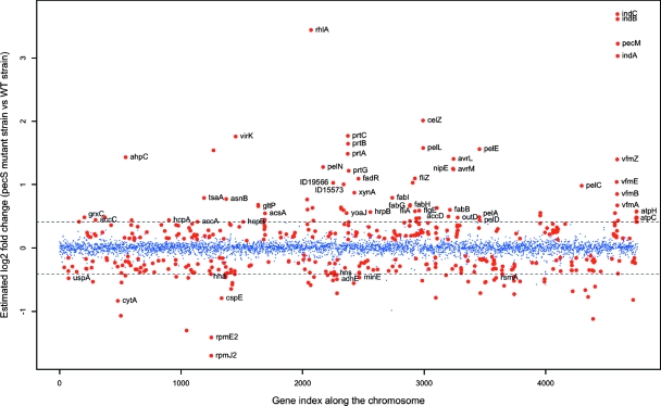
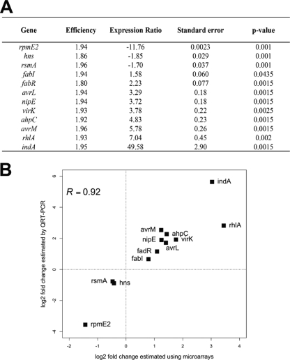
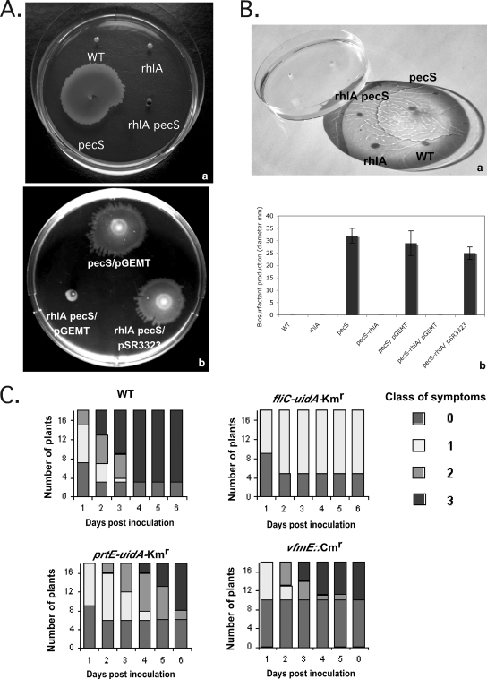
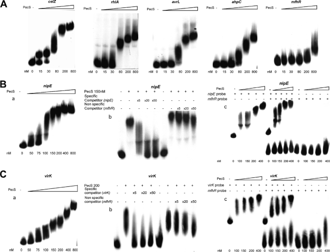
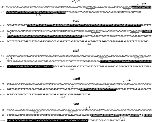
Pathogenicity of the enterobacterium Erwinia chrysanthemi (Dickeya dadantii), the causative agent of soft-rot disease in many plants, is a complex process involving several factors whose production is subject to temporal regulation during infection. PecS is a transcriptional regulator that controls production of various virulence factors. Here, we used microarray analysis to define the PecS regulon and demonstrated that PecS notably regulates a wide range of genes that could be linked to pathogenicity and to a group of genes concerned with evading host defenses. Among the targets are the genes encoding plant cell wall-degrading enzymes and secretion systems and the genes involved in flagellar biosynthesis, biosurfactant production, and the oxidative stress response, as well as genes encoding toxin-like factors such as NipE and hemolysin-coregulated proteins. In vitro experiments demonstrated that PecS interacts with the regulatory regions of five new targets: an oxidative stress response gene (ahpC), a biosurfactant synthesis gene (rhlA), and genes encoding exported proteins related to other plant-associated bacterial proteins (nipE, virK, and avrL). The pecS mutant provokes symptoms more rapidly and with more efficiency than the wild-type strain, indicating that PecS plays a critical role in the switch from the asymptomatic phase to the symptomatic phase. Based on this, we propose that the temporal regulation of the different groups of genes required for the asymptomatic phase and the symptomatic phase is, in part, the result of a gradual modulation of PecS activity triggered during infection in response to changes in environmental conditions emerging from the interaction between both partners.


Figures

FIG. 1.
FIG. 1.
Expression ratios of genes in pecS mutant versus wild-type strain along the chromosome. Blue points represent genes that were expressed similarly in pecS and wild-type (WT) strains. Red points represent genes that were differentially expressed in the pecS mutant versus the wild-type strain with an FDR-adjusted P value of <0.05. Genes with a greater than ±1.33-fold difference in the expression ratio fall outside of the field marked by dotted lines. The gene index is organized so that gene 1 corresponds to the origin of replication, OriC.
FIG. 2.
FIG. 2.
Validation of microarray results by qRT-PCR. (A) Expression ratios of genes in pecS mutant versus the wild-type strain measured by qRT-PCR. Expression of each gene was normalized to the expression of the two housekeeping genes, rpoA and ffh. A positive expression ratio indicates upregulated genes in the pecS background, and a negative expression ratio indicates downregulated genes in the pecS background. Standard errors were calculated from the data from three independent biological replicates. (B) Comparison of gene expression measurements by microarray hybridization and real-time qRT PCR. The correlation coefficient (R) is given.
FIG. 3.
FIG. 3.
Phenotypic analysis. (A) Swarming motility of various bacterial strains (a). Bacterial strains were inoculated onto LB-1% agar plates and incubated at 30°C for 24 h. Swarming motility of the rhlA pecS double mutant complemented with plasmid pSR3323 expressing RhlA is shown (b). (B) Biosurfactant production of various bacterial strains. Bacterial strains were inoculated by stabbing 0.7% semisolid agar plates and incubated at 30°C for 8 h. A wet puddle appears on the surface of the agar medium for the pecS strain producing biosurfactant (a). The size of the halo of this secreted substance is a direct assay of the surfactant production (b). Data shown are the means ± standard deviations of at least three independent experiments. (C) Interactions between the fliC, prtE, and vfmE mutants and S. ionantha plants. Infection was performed on a single leaf per plant by infiltration of 107 bacteria in Saintpaulia. Symptom occurrence was scored daily for a week. Symptoms are classified in four stages: stage 0, no symptoms; stage 1, rotting confined to the infiltrated zone; stage 2, maceration of the leaf limb; stage 3, maceration of the whole leaf including the petiole. At least three independent experiments were performed, and for each, mutant scores were different from wild-type scores, with a P of < 0.05. Results of a typical experiment are presented. Results obtained with the prtE-uidA-Kmr and vfmE::Cmr mutants were confirmed with independent mutants (prtE::MudII1734 and vfmE-uidA-Kmr). For the fliC-uidA-Kmr mutant, two independent transductants were analyzed, and similar results were obtained. WT, wild type.
FIG. 4.
FIG. 4.
Band shift assay for PecS-DNA binding. (A) Labeled DNA probes, corresponding to promoter regions of rhlA, avrL, and ahpC, were incubated with increasing concentrations of PecS, as indicated at the bottom. The celZ regulatory region was used as a positive control (44), and the mfhR nonspecific fragment derived from the coding region of a transcriptional regulator not controlled by PecS was used as a negative control. (B) Specificity of PecS binding on nipE regulatory region. EMSA shows binding of PecS to the 400-bp nipE regulatory region (region −300 to +100 relative to the translation start site) with an increasing PecS concentration (0, 50, 75, 100, 150, 200, 400, and 800 nM) in the presence of 30 fmol of radiolabeled probe (a). In the reactions mixtures containing 30 fmol of radiolabeled probe and 150 nM PecS, competition assays were performed with the molar excesses of the unlabeled 400-bp nipE regulatory region specific competitor indicated above the lanes (×0, ×5, ×20, and ×50) and with the indicated molar excesses of unlabeled nonspecific competitor DNA derived from a 250-bp fragment of the mfhR coding region (b). Simultaneous titrations were performed with PecS and both 32P-labeled nipE probe and 32P-labeled mfhR probe (c). (C) Specificity of PecS binding on virK regulatory region. EMSA shows binding of PecS to the 440-bp virK regulatory region (region −370 to +70 relative to the translation start site) with an increasing PecS concentration (0, 50, 75, 100, 150, 200, 400, and 800 nM) in the presence of 30 fmol of radiolabeled probe (a). In the reaction mixtures containing 30 fmol of radiolabeled probe and 200 nM PecS, competition assays were performed with molar excesses of the unlabeled 440-bp virK regulatory region specific competitor indicated above the lanes (×0, ×5, ×20, and ×50) and with the indicated molar excesses of unlabeled nonspecific competitor DNA derived from a 250-bp fragment of the mfhR coding region (b). Simultaneous titrations were performed with PecS and both 32P-labeled virK probe and 32P-labeled mfhR probe (c).
FIG. 5.
FIG. 5.
Sequence of the ahpC, avrL, rhlA, nipE, and virK regulatory regions. The transcription start sites are indicated by arrows and by a bold character, and the −10, −35 RNA polymerase-binding regions are underlined. The initiation codons are in bold letters and are underlined. The PecS binding sites are boxed in black. The Shine-Dalgarno sequence (S.D.) corresponding to the ribosome binding sites are underlined.

Similar articles

Cited by

References

    1. Andrews, J. 2001. Determination of minimum inhibitory concentrations J. Antimicrob. Chemother. 485-16. - PubMed
    1. Bardonnet, N., and C. Blanco. 1992. ′uidA-antibiotic-resistance cassettes for insertion mutagenesis, gene fusions and genetic constructions. FEMS Microbiol. Lett. 72243-248. - PubMed
    1. Beaulieu, C., M. Boccara, and F. Vangijsegem. 1993. Pathogenic behavior of pectinase-defective Erwinia chrysanthemi mutants on different plants. Mol. Plant-Microbe Interact. 6197-202.
    1. Benjamini, Y., and Y. Hochberg. 1995. Controlling the false discovery rate: a practical and powerful approach to multiple testing. J. Royal Stat. Soc. Ser. B 57289-300.
    1. Boccara, M., A. Diolez, M. Rouve, and A. Kotoujansky. 1988. The role of the individual pectate lyases of Erwinia chrysanthemi strain 3937 in pathogenicity on Saintpaulia plants. Physiol. Mol. Plant Pathol. 3395-104.

Publication types

MeSH terms

Substances